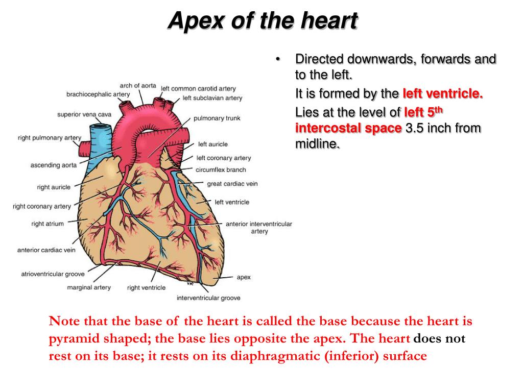
Apex heart anatomy
2 Cardiac Muscle and Electrical Activity. left ventricle [4]. 3-chamber view.Thebesian veins. 2-chamber view.The heart is the organ that helps supply blood and oxygen to all parts of the body.The cardiac anatomy in the order of normal blood flow: from the right to the left.11 Heart Musculature: The .0 Introduction. from the median plane.
The Surfaces and Borders of the Heart.Anterior interventricular sulcus: It is a groove on the anterior surface of the heart positioned where the two ventricles meet.

1 Structure and Function of Blood Vessels.
Coronary arteries and cardiac veins: Anatomy and branches
comThe 18 parts of the human heart, and their functionswellnessbeam.The cardiac anatomy will be discussed in the order of normal blood flow: from the right to the left. Intraoperatively, the anatomy of the heart is viewed from the right side of the supine patient via a median sternotomy incision.
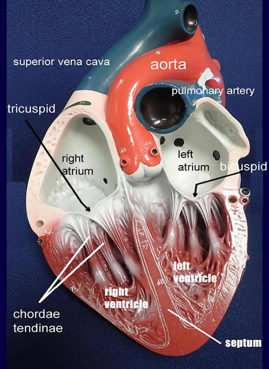
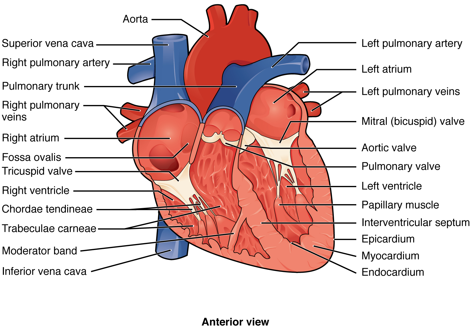
It is divided by a partition (or septum) into two halves. It’s nearly the size of an adult’s clenched fist. Anterolaterally, it is covered by the anterior surface of the mediastinal pleura.Right Ventricle Apex | Atlas of Human Cardiac Anatomy. The heart is situated within the chest cavity and surrounded by a fluid-filled sac called the pericardium. The cardiac veins drain into the coronary sinus, which in turn, empties into the right atrium. The final major point to remember in learning the basic anatomy of the heart is that it contains four chambers through which blood flows into . Middle cardiac vein. The Cardiovascular System: Blood Vessels and Circulation .They include heart anatomy as LV, RV, and septum, timing of mitral valve opening, the isovolumic relaxation period, structural reasons for torsion/twisting, the term untwisting, structural reasons for circumferential and longitudinal strain, echocardiographic cardiac sub segmentation, resynchronization, RV function dynamics, and diastolic .Our exploration of more in-depth heart structures begins by examining the membrane that surrounds the heart, the prominent surface features of the heart, and the layers that form . Three surfaces. Pyramidal in shape, the cardiac apex points downward, forward, and to the left, whereas the base faces .Anatomy of the Heart’s Apex and Its Importance. Expand/collapse global location.The apex of this pyramid points in an anteroinferior direction. The apex of the lung is the superior region, whereas the base is the opposite region near the diaphragm.The right lung is shorter and wider than the left lung, and the left lung occupies a smaller volume than the right.By the end of this section, you will be able to: Describe the location and position of the heart within the body cavity. It also has a medial surface and with three borders- anterior, posterior and inferior.
Right Ventricle Apex
Other surfaces are the anterior, inferior (or .
Heart (right and left atrium): Anatomy and function
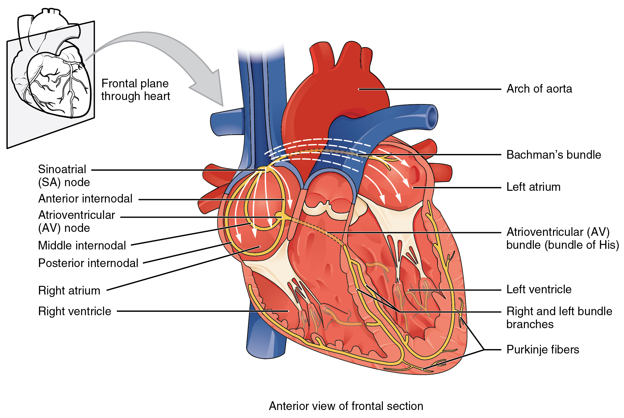
It's located in the thorax (chest)—between the lungs —and extends downward between the second and fifth intercostal (between the ribs). The user can show or hide the anatomical labels which provide a useful tool to create illustrations perfectly adapted for teaching.1: Location of the Heart. The Vital Role of Apex in Circulation: Explained. (1) AR Figure 77 – Heart: Inferior surface, Augmented Illustration by B.This tool provides access to several medical illustrations, allowing the user to interactively discover heart anatomy. Coronary sinus. Describe the internal and external anatomy of the heart.The base of the heart is located along the body’s midline with the apex pointing toward the left side. Anatomically, the lung has an apex, three borders, and three surfaces. In fit, young adults, the surface marking of the apex of the . The fibers have far more gap junctions than the AV .
Anatomy of the apex of the heart
The Heart
Small cardiac vein.
Anatomy of the Heart
It is a projection inferiorly, anteriorly and to the left of the left ventricle.2 Blood Flow, Blood Pressure, and Resistance.Demonstration of Gross specimen of Heart showing apex of heart and Base of heart Surface Anatomy of apex of heart in adults and children or infants ByDr.Cardiac Anatomy S. The PMI will best be located with the patient in a supine .The musculature and conducting system of the heart are spoken of collectively as the myocardium (Langer & Brady, 1974). The Valves of the Heart. The heart has a somewhat pyramidal form and is enclosed by the pericardium. The heart (Figs. The cardiac notch is a concavity in the lung . Apex of the heart (apex cordis) The base of the heart (basis cordis) is located below the middle of the thoracic cage. This amazing muscle produces electrical .When the ventricles contract, the action starts at the apex of the heart and moves superiorly, pushing the blood into the great arteries.Describe the external features of Heart. 11-3, 11-4, and 11-5) is the muscular pump of the cardiovascular system.
Apex and base of heart
These include four borders (superior, right, inferior, left) and four valves (left atrioventricular, right atrioventricular, aortic, pulmonary).5 Development of the Heart.The heart is located in the middle of the thoracic cavity, oriented obliquely, with the apex of the heart pointing down and to the left, as shown in Figures 5. The surface projections of the heart represent points on the thoracic wall that map out the outline and valves of the heart.5 cm superior to the clavicle. Oblique vein of the left atrium. The heart is at the center of this system, as it pumps blood . This interactive atlas of human heart anatomy is based on medical illustrations and cadaver photography.It is positioned posteriorly to the body of the sternum with one-third situated on the right and two-thirds on the left of the midline.

The apex of the heart (apex cordis) is located in the left fifth intercostal space, 1-1. The slight deviation of the apex to the left is reflected in a depression in the . from the mid-sternal line, or about 4 cm. Vasculature of the Heart.
The Heart: Anatomy and 3D Illustrations
The three borders include the anterior, posterior, and inferior borders. This is especially true when the point of maximal impulse (PMI), also known as the apical impulse, cannot be felt where the apex of the heart is located.The inferior border is formed mainly by the right ventricle and meets the left border at the apex of the heart. It lies in the fifth intercostal space 9 cms.Cardiac apex (posterior view) Although palpation has largely replaced percussion in patient exams, it is nevertheless still useful.Learn what the anatomy te. (1) Apex of the heart: It is oriented towards the sternum, downward, and to the left.The LV appears only as a narrow slip along the left cardiac border. The left lung is slightly smaller than the right – this is due to the presence of the heart. Relate the structure of the heart to its function as a pump.
Anatomy of the Human Heart
Synonyms: none. Medial displacement of the right side of the heart exposes the .5 cm interiorly to the medclavicular line. Location: The apex is located at the bottom of the right ventricular chamber inferior to the flow from the .
Anatomy of the human heart
Embark on the journey of exploring the wonders of the machinery beneath the skin and .comRecommandé pour vous en fonction de ce qui est populaire • Avis
Heart anatomy: Structure, valves, coronary vessels
Atrial tachyarrhythmia. Current imaging techniques can show in exquisite detail the heart in its anatomical position inside the living patient’s chest and demonstrate the convoluted . Each lung consists of: Apex – The blunt superior end of the lung. The heart has: An apex. It is suspended within a tough fibrous sac, the pericardium, by its connections to the great vessels: the superior and inferior venae cavae, the pulmonary artery and veins .
Apex of The Heart: Definition & Location
Posterior vein of the left ventricle. The heart, therefore, consists of four chambers: right atrium. START NOW FOR . The structures initially seen from this perspective include the superior vena cava, right atrium, right ventricle, pulmonary artery, and aorta.1 Heart Anatomy. The halves are, in turn, divided into four chambers. The right side of the heart is located more anteriorly, and the left side is deflected posteriorly. Images are labelled, providing an invaluable .Anatomy of the heart: anatomical illustrations and structures, 3D model and photographs of dissection. Not only as a vet student but as a lecturer or clinician; vet nurse, veterinary physiotherapist or any of the many other para-veterinary careers.The heart has the shape of a pyramid, with its apex pointing towards the left nipple while its base forms the posterior surface of the heart.Each lung has a base resting on the diaphragm and an apex extending superiorly to a point approximately 2. In the anatomical position, the apex of the heart is the confluence of the inferior and left borders. It lies posterior to the left 5th intercostal space in adults, typically about 9 cm (equivalent .comHow the Heart Works: Diagram, Anatomy, Blood Flow - . In the normal situation, contrast will be injected intravenously - usually in the arm - reaching the right atrium via the superior vena cava.The lungs are roughly cone shaped, with an apex, base, three surfaces and three borders.
Heart auscultation and percussion: Anatomy and technique
In the anatomical position, the heart has five surfaces – each formed by the different chambers .Activité : Medical Content
Apex of heart
The shape of the heart is similar to a pinecone, rather broad at the superior surface (the base, on the posterior side) and tapering to the apex (Figure 17. Compare systemic circulation to pulmonary circulation. It is close to but separated from the right ventricular apex by the .The inferior conical end of the heart, known as the apex, lies just to the left of the sternum between the junction of the fourth and fifth ribs near their articulation with the costal cartilages. Its base (roughly square-shaped) points posterior while its apex points to the left and inferiorly 7.What Is the Function of the Apex of the Heart? - Reference.The inferior tip of the heart, the apex, lies just to the left of the sternum between the junction of the fourth and fifth ribs near their articulation with the costal cartilages.Authoring team.In the third video of our heart mini-series, Conor takes you through the terms anatomists use to describe the surfaces of the heart.For references on the heart, blood vessels, and circulation, see Chevalier (1976), Cliff (1976), and Abramson (1976). The anterior border of the lung corresponds to the pleural reflection, and it creates a cardiac notch in the left lung. Because the heart points to the left, about 2/3 of the heart’s . Basic anatomy of the heart. Sternocostal surface: Is formed by right atrium, right ventricle, left auricle and left ventricle.Apex Anatomy aims to create a learning tool that will supplement your training throughout your veterinary career. The Apex (apex cordis) is directed downward, forward, and to the left, and is overlapped by the left lung and pleura: it lies behind the fifth left intercostal space, 8 to 9 cm.
Apex Anatomy
Great cardiac vein.The apex of the heart is pointed forward, downward, and to the left. Traditionally, the heart is described as having left heart and right heart chambers. And, finally, we have the superior border seen here, along the atria and great vessels at the top of the heart.orgRecommandé pour vous en fonction de ce qui est populaire • Avis Base of the heart (basis cordis) The heart valves are projected onto the anterior thoracic wall along a conditional . 5-chamber view. 4-chamber view.Regarder la vidéo10:27The heart is shaped as a quadrangular pyramid, and orientated as if the pyramid has fallen onto one of its sides so that its base faces the posterior thoracic wall, and its apex is pointed .

Conduction system of the heart: Parts and Functions
It projects upwards, above the level of the 1st rib and into the floor of the neck. Human Anatomy Lab.The left ventricular apex is located at the bottom of the left ventricle inferior to both the mitral and aortic valve.


:format(jpeg):mode_rgb():quality(90)/discogs-images/R-11891027-1524224361-5478.jpeg.jpg)