Bacteria cell simple diagram
Bacteria cell: the bacterial cell wall.0 µm in length. File usage on Commons.semanticscholar. The cell membrane surrounds a cell’s cytoplasm, which is a jelly-like substance containing the cell’s parts.comBacterial cell structure - Buy Royalty Free 3D model by Eberssketchfab.Bacteria is a unicellular prokaryotic organism.
Bacterial cell structure
Diagram shows the process how bacterial cells experiencing poor growth conditions can form endospores in the following steps: 1.govThe Atlas of Bacterial & Archaeal Cell Structure: an .Bacterial cells have all of the organelles that plant cells do, except for a true nucleus, chloroplasts or mitochondria. How Big is a Bacteria. Among the smallest bacteria are members of the .
Structure of Bacterial Cell (With Diagram)
4: Colony and Cell Morphology; Simple Stains
Overview
Bacterial cells
The cytoplasm fills the interior of the cells, and bathes the nucleoid. Bacteria do not grow during the lag phase. Bacteria cells are typically 0. It includes two domains – bacteria and archaea. The cells are all. Unlike the eukaryotic (true) cells, bacteria do not have a membrane enclosed nucleus.The bacteria diagram given below represents the structure of a typical bacterial cell with its different parts. in plants and animals. Size of this PNG preview of this SVG file: 377 × 599 pixels. However, for other Bacteria, it makes up only about 10% of the cell wall (Fig. Illustration of the bacteria cell structure. DNA is replicated, 2. A prokaryotic cell is a type of cell that does not have a true nucleus or membrane-bound organelles. It is 20–30 μm in diameter and 15 μm in length. Outer Membrane. cellular division of cytoplasmic membrane, 3. The cell envelope encases the cytoplasm and all its components. It is single-stranded, unlike DNA in a nucleus which is . All living organisms are classified into three major domains: Domain Eukaryota (), Domain Eubacteria (true bacteria), and Domain Archaea (archaebacteria). We hope it will be a useful tool for microbiology courses, serving as a quick introduction to the cells and what they contain before students go on to study .Last Updated: October 4, 2019.It is a gel-like matrix composed of water, enzymes, nutrients, wastes, and gases and contains cell structures such as ribosomes, a chromosome, and plasmids.3, so we will not go over that in detail here . Organisms within the domains . Bacteria Poster.Figure 5: (A) Endospores form from bacterial cells. Some bacteria have flagella.The bacteria life cycle consists of the lag phase, the log or exponential phase, the stationary phase and the death phase. The cytoplasm is where most of the cell’s chemical reactions take place.English: A simple diagram of a yeast cell, labelled in English.For some Bacteria, peptidoglycan makes up as much as 90% of the cell wall. Parts of a Cell Cut and Stick Worksheet. References and Sources: Bacteria are unicellular organisms with a simple structure. These two groups of Bacteria can be distinguished based on a staining procedure. spore coat formation begins, 6.000039 inch), with a total mass of 10 −14 gram—equal to that of 8,000,000,000 hydrogen atoms.Domain Eubacteria includes the true bacteria. It is in between the cell membrane and the capsule/slime layer. Bacterial cell diagram showing the basic organelles of a bacterium. Viruses PowerPoint. The cell wall, plasmid, cytoplasm and flagella are clearly marked in the .2 μm in diameter (1μm = about 0. cocci) – a coccus is a . It shows the cytoplasm, nucleus, cell membrane, cell wall, mitochondria, and vacuole.
Ultrastructure of a Bacterial cell
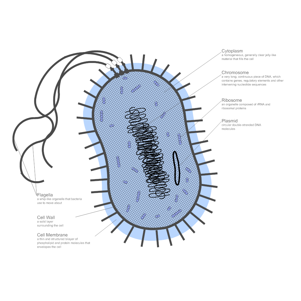
Bacteria are all single-celled.It shows the cytoplasm, nucleoid, cell membrane, cell wall, mitochondria (which bacteria lack), plasmids, flagella, and cell capsule.3 µm, while a few others are so big that they are visible even to the naked eye.Figure 1: Bacterial cell – diagram with label.
They have an outer cell wall that gives them shape. maturation of . The cell wall of the bacteria maintains the shape of . Other resolutions: 151 × 240 pixels | 302 × 480 pixels | 483 × 768 pixels | 644 × 1,024 pixels | 1,288 × 2,048 pixels | 390 × 620 pixels.comRecommandé pour vous en fonction de ce qui est populaire • Avis
Bacteria Diagram- Simple Structure with Labels, Function
Key points to note when comparing and contrasting the structure of bacterial cells with animal and plant cells are that they: Embryonic stem cells close . Flagella are whip-like structures used for movement.In this educational video, we provide a quick and easy tutorial on sketching a bacterial cell diagram and labeling its organelles.Microscopy and cells (CCEA) Bacterial cells. In addition to the nucleus, eukaryotic cells .
Cell structure Structure of fungal and bacterial cells
More Science resources Bacteria Labelled Diagram Vaccine PowerPoint Periodic Table of Chemical Elements . The cell's DNA is extensively folded to form a body called the nucleoid.Overview of prokaryotes (bacteria and archaea). Bacteria lack a .

Flagella are used for locomotion.Bacteria are single-celled organisms. Structural features of prokaryotic cells.


And so, inspired by the atlases of eukaryotic cell structure from the 1960s, here we offer an atlas of bacterial and archaeal cell structure, highlighting many of the molecular machines we have discovered so far. Bacteria Definition. mitochondria and chloroplasts of eukaryotic cells.The nucleoid and some other frequently seen features of prokaryotes are shown in the diagram below of a cut-away of a rod-shaped bacterium.Simple diagram of bacterium (blank).
The Atlas of Bacterial & Archaeal Cell Structure
Français : Schéma d'une cellule de levure avec des étiquettes en anglais. By this method, cells are exposed to a stain, crystal violet, that binds to peptidoglycan. Types of Germ PowerPoint. Simple Diagram of the Heart Labelling Activity. However, they do adjust to their environment and metabolize, that is, produce vitamins and amino . Just under the rigid cell wall is the more fluid cell membrane. To examine cells and learn more about their structure, we need to be able to see them in very fine detail. Español: Diagrama de una célula de levadura con etiquetas en Inglés.
Molecular Expressions Cell Biology: Bacteria Cell Structure
The cell membrane surrounds a cell’s cytoplasm, .For example, Thiomargarita namibiensis is the largest and longest bacteria with a diameter .Despite the tremendous genetic diversity seen in prokaryotes, cell morphology falls into just a few categories. Prokaryotes are free-living and photosynthetic (produce their food), parasitic (living inside other . The ribosomes present in mitochondria and chloroplasts are smaller than 80S . The flagella of Eukaryotic cells contain 9+2 microtubles but each flagellum in bacteria is made up of a single fibril. prespore formation begins, 4. cortex forms, 5.comCELLS alive! Going Offlinecellsalive. It is the largest domain that includes a large group of organisms.Bacteria, microscopic single-celled organisms that inhabit virtually all environments on Earth, including the bodies of multicellular animals.How Big is a Bacteria. Membrane Proteins.org3D design Bacteria cell model - Tinkercadtinkercad. bacterium) are unicellular prokaryotic microorganisms which divide by binary fission. Typical examples include: coccus (circle or spherical); bacillus (rod-like); coccobacillus (between a sphere and a rod); spiral (corkscrew-like); filamentous (elongated); Cell shape is generally characteristic of . Each organelle carries out a specific function in the cell. Sedimentation coefficient: 80S ; Molecular weight: 40 × 10 6 daltons.Bacterial cells contain a cell membrane and a cell wall, which provides structure. Free Chromosomal DNA – Bacterial cells have DNA free in the cytoplasm. Date: 18 January 2016: Source : Own work: .Critiques : 4
Cell
Storage granules contain a reserve of nutrients - typically polymeric forms of b -hydroxybutyrate and phosphate. _Image credit: modified from .
Interactive Bacteria Cell Model
Labelled diagram of a bacterial cell: Cell Capsule: It is a slime layer composed of a thick polysaccharide.
Cell Structure
Prokaryotic cells (article)
Cell wall The bacteria’s cell wall is the outer rigid and chemically complex structure. Cell wall: Cell walls of bacteria are . Français : Un schéma d'une . The cytoplasm enclosed within the cell membrane does not exhibit much .The cell structure of a bacterial cell consists of a complex membrane and membrane-bound protoplast.All cells have a cell membrane that separates the inside and the outside of the cell, and controls what goes in and comes out. Key points: Prokaryotes are single-celled organisms belonging to the domains Bacteria . Cell wall – The cell wall is not made of cellulose, but is instead made of peptidoglycan. This means they do not have a nucleus or any other structures. Cytoplasm is a jelly-like fluid that fills the cell.Bacteria cell anatomy.prokaryotic cells of the blue-green algae and bacteria. Prokaryotes are simple, single-celled organisms that are the most primitive life form on earth. Vector bacterial cell anatomy isolated on white background.Human cells, the virus infects cells. Bacteria (sing.What is a Prokaryotic Cell. There are also cell walls, cytoplasm, and nucleoids . Plasmids are small rings . Bacterial cells form cocci (round), bacillus (rod), and curved structures.In this article we will discuss about the Structure of Bacterial Cell.Bacterial Identification Virtual Lab - HHMI BioInteractivebiointeractive. Bacteria have cell walls located outside of their cell membrane. Vector - Plant Cell.

From Wikimedia Commons, the free media repository.Bacteria (Prokaryotes) are simple in structure, with no recognizable organelles.4 Bacteria: Cell Walls.Last Updated: November 6, 2020.3: Eukaryotic Cell: Structure and Function is shared under a not declared license and was authored, remixed, and/or curated by LibreTexts. Vector diagram.The smallest known cells are a group of tiny bacteria called mycoplasmas; some of these single-celled organisms are spheres as small as 0. Cytoplasmic Membrane.1: General bacterium diagram.Bacteria tend to display the most representative cell morphologies, with the most common examples listed here: Bacterial Cell Morphologies. They are considered prokaryotes because of the .orgBacteria Model | College of DuPage Librarycodlrc. 3d illustration bacteria cell stock pictures, royalty-free photos & images Outbreak of Chinese influenza - called a Coronavirus or 2019-nCoV, Group of blue-colored cells under microscope High quality 3d render of cells bacteria cell stock pictures, royalty-free photos & images
ᐉ Bacterial Cells: Types, Differences, Diagrams, and more!
Gram-negative Cell Wall. Detailed diagrams and descriptions of bacterial cell morphology can be found in OpenStax Microbiology, Chapter 3. They are found in the eukaryotic cells i. Having said that though, it is also important to note that most bacteria (about 90%) have a cell . Plant Cell Diagram Worksheets.Structure and Physical Characteristics.
Bacteria: Cell Walls
Cell morphology Bacteria come in a wide variety of shapes. Transformation, in molecular biology and genetics, is the genetic modification of a cell caused by the direct absorption and incorporation of exogenous genetic material from its environment through the cell membrane (s). have a more simple structure compared to animal, plant and fungal cells and are usually much smaller.

The cell wall is made up of sugar (glycan) chains connected by .