Best antibody concentration for flow

Their purpose is to ensure the observed staining is due to specific . Exclusive positivity on the y-axis (upper left quadrant) and the x-axis (lower right quadrant) shows cells stained for MAP2 and CD24, .What is the cell biology and expected experimental outcome? Add 2 mL of Flow Cytometry Staining Buffer and centrifuge at 400-6—x g for 4-5 minutes at room temperature.
Antibody Titration for Flow Cytometry
The optimal antibody for capture vs.Titration for flow cytometry is used to determine the antibody amount and concentration resulting in the highest signal of the positive population along with the lowest signal of . Results are best when the total amount of antibody for all samples is prepared as a .
We can also browse the antibody conjugates available on the CST website, including antibody conjugates that are validated for flow cytometry experiments.Check out our Antibody Staining Guide for Flow Cytometry to determine how to best combine antibodies binding extracellular and intracellular targets.
Manquant :
flowWhat to Know About the COVID-19 Antibody Test
When using an antibody for the first time, you may need to optimize its dilution for your specific application and experimental setup.Dilute the primary and secondary antibodies in the suspension buffer.After designing a multicolor flow cytometry panel and securing the necessary cells and reagents, the process of optimization of the panel can begin. Different fluorescent conjugates are presented in descending order, 405 nm .During production of concentrated monoclonal antibody formulations by tangential flow ultrafiltration (TFF), high viscosities and aggregation often cause extensive membrane fouling, flux decay and low product yields. For instance, . Place on ice, in the dark, for 20 minutes.Each antibody consists of four polypeptides – two heavy chains and two light chains.That testing is for assessing antibody levels against SARS-CoV-2, the virus that causes COVID-19.
Flow cytometry recommended controls
Titration of Antibodies
Manquant :
flow Incubate plate at 5% CO 2 at 37°C for 2 hours. Right, immunofluorescent staining of human cell line U-251 MG. In the case of AIM2, one of articles we reviewed used BioLegend 652803 at 1:20 for B-lymphocyte flow cytometry staining.Antibody dilutions and titer
KLH) of the same antibody subclass, with the same conjugated fluorophore, that is obtained from the same supplier as your test antibody and used at the same concentration.
Titrating Antibodies for Flow Cytometry
Determine the optimum antibody concentrations for both the capture and detection antibody. We hope this is helpful. What are antibodies and what do they do? Antibodies are large ‘Y’ shaped .The correct antibody concentration is very important to ensure best resolution and consistent results in flow cytometry analysis. Remember isotype controls should not be used to determine compensation levels or the negative population.Figure 1: Determining the appropriate staining protocol path for the CD4 (RM4-5) and TCRβ (H57-597) example, using the Antibody Staining Guide for Flow Cytometry .
Titrating antibodies for flow cytometry is a useful step in effective and economic panel design.When acquiring data on the flow cytometer, be sure that the stained cells are on scale; look at the tube with the highest concentration of antibody for each .The optimal concentration must be re-evaluated every time a new primary or secondary antibody is used, or when experimental conditions change.
Whole Blood Staining Protocol for Flow Cytometry Analysis
Like other immunoassays, the primary antibody for your target should be tested and validated on Simple Western.In this guide, antibody scientists share what we’ve learned about getting the best possible image from your IF-ICC experiments. Search Antibodies.If antibody-based protein evaluation is performed in a quantitative manner, signal-to-noise ratio and dynamic range are two of the most critical objective parameters . The objective of titrating fluorochrome-labeled antibodies is to identify the optimal concentration for a given marker-fluorochrome pair that results in .Optimizing Flow Cytometry Experiments – Part 2 How To Block Samples (Sample Blocking) Written by Tim Bushnell, PhD.
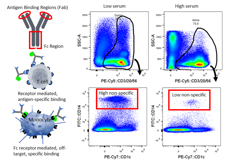
Coat the activated T cell wells with the anti-CD3 antibody by diluting the anti-CD3 antibody at 1 µg/mL in sterile PBS. Most antibodies come with a recommended concentration from the .You can have a look at our Validated Antibody Database antibody reviews.Principles of the Flow Cytometer. Figure 1: Immunocytochemistry (ICC) locates proteins associated with neuronal nuclei, soma, and axons (left). The formula used is: Concentration (stock) × Volume (stock) = Concentration (dilute) × Volume (dilute) The stock concentration of your antibody: /.It is recommended to test 0. For example, a . The concentration of antibody required: /. Next to the flow cytometer itself, the most important component of a flow cytometry experiment comes down to the antibodies. Intracellular staining can be affected by binding of both antibody and fluorophore to intracellular components, therefore choice of fluorophore and extra controls may be necessary.The correct isotype control is an antibody generated against an irrelevant antigen (e. The achievements of cytometry moved the field to a .The objective of titrating fluorochrome-labeled antibodies is to identify the optimal concentration for a given marker-fluorochrome pair that results in the best possible separation between the positive and negative cell populations, while minimizing the background within the negative population. Even antibodies validated for other applications such as FACS or IHC may work on Simple Western. In my previous blog on experimental optimization, we discussed the idea of identifying the best antibody concentration for staining the cells. This webpage is a useful resource for researchers and students .
Flow cytometry protocol
Results are best when the total amount of antibody for all samples is prepared as a single master stock at a concentration such that no less than 1.If using a viability dye, make sure to include a single stained control for viability (for compensation) 3. The first step in that optimization is titration of your antibodies. detection can only be . This wash step can be repeated.The goal is to develop a basic working method by determining the antibody which should be the capture antibody and which antibody should be the detection antibody.Cell Viability
Antibody Titration
5 μg depending on antibody). In this process, following a standard protocol to be used in the final analysis, you stain a known amount of cells with decreasing . Prepare the concentration so that 10 l can be a dded to each sample. Isotype controls can be conjugated or unconjugated, and should match the host species, the Ig subclass, and conjugation of the primary antibody used against the target of interest. “Monoclonals, especially .
Optimizing Flow Cytometry Experiments
This allows you to determine what concentration of antibody leads to .Flow cytometry (FACS) staining protocol (Cell surface staining) is a webpage that provides detailed instructions on how to prepare and stain cells for flow cytometry analysis. Discard supernatant.Written by Tim Bushnell, PhD.
Antibody Titration in Flow Cytometry
We recommend testing antibody dilutions from 1:50 to 1:100 initially.

According to their functional characteristics, antibodies can be subdivided into variable and constant regions. Prepare complete RPMI 1640 medium by supplementing RPMI 1640 medium with fetal bovine serum to a final concentration of 10% and . Add the antibody cocktail to a 100 μL aliquot of whole blood. We did this through a process called titration, which focuses on finding the best . The basis of flow cytometry is the measurement of how light is scattered in the forward or side direction as it passes through a particle. The variable region, made up of the N-terminal ends of the heavy and light chains, contains the antigen binding site.To perform a simple antibody titration, start with the manufacturer’s recommended concentration, perform serial 2-fold dilutions, and plot the stain index (SI), which is a . Too little antibody and the specific positive signal remains weak and susceptible to small changes in staining conditions, too much antibody and the unspecific background signal starts to increase (Fig.Simple Western is an open platform so any antibody may be used for detection, including Western blot antibodies.Antibody titration — excess antibody can bind with low affinity, creating background fluorescence that can reduce signal resolution. Incubate for 30 minutes to 1 hour at 2–8°C with rotation.To address these challenges, the co-solutes histidine or imidazole were added at high concentrations from 250 to 320 mM to . It covers topics such as cell harvesting, washing, concentration, antibody labeling, fixation, and data acquisition.Isotype controls have been optimized for surface staining.
Immunoassay Methods
An isotype control is an antibody selected as a negative control for flow cytometry experiments, and is used to detect non-specific binding.Flow cytometry predominantly uses direct staining with conjugated primary antibodies to detect markers of interest, especially when multiplexing for immunophenotyping, however, not all antibodies are available with a fluorescent marker or one that is compatible with other fluorophores or fluorescent proteins.
Dilution Calculator
Add the antibody to the cells and gently mix.
Simple Western Frequently Asked Questions
Best practices in flow cytometry dictate that . Add diluted antibody to the 3 wells at 2 mL/well. Hence, the volumes of antibodies used in multiparametric flow cytometry profoundly affect the cost of experimentation. Titration is an essential . The concentration that shows the .Popular answers (1) For immunophenotyping studies, it is always good to keep cell numbers less than 10,000/uL to have a good flow of cells besides having enough cells for the antibody.5 mg/mL of human IgG, no erythrophagocytosis was induced by NILK-2401, up to the highest antibody concentration tested (300 μg/mL).to find the optimal concentration of antibody that results in the brightest signal of the positive populationwhile avoiding backgroundstaining, thus maximizing .Antibody titration allows you to optimize the concentration of antibody used by determination of the best signal to noise ratio, or stain index.