Chest x ray radiology
Significant pectus excavatum has an index greater than 3.What is a chest X-ray? The chest x-ray is the most commonly performed diagnostic x-ray examination.
Assessment of pulmonary hila on chest x-ray (approach)
The chest X-ray is the most frequently requested X-ray at the radiology department. This index uses the vertebral body diameter as a .comHow To Read A Chest X-ray [Step-by-Step] - TheMDJourneythemdjourney.Find a Chest X-ray near me & book an appointment online for free.FREE download PDF Word format X rays Chest PA View . Robin Smithuis and Otto van Delden. Radiology and You.
Manquant :
chest x rayThe Chest X-Ray: A Systematic Teaching Atlas
The underlying cause (such as a lung tumor or pleural effusion) may also be visible.
A radiologic technologist will guide you through the process. with severe LV enlargement the short axis .Chest X-Ray - Basic Interpretation. & Canada: 1-877-776-2636 Outside U. A chest x-ray produces images of the heart, lungs, airways, blood vessels .Balises :Chest RadiologyThe Chest X-Ray
Chest X-ray Interpretation
Doctors typically use this procedure to help diagnose breathing difficulties, a bad or persistent cough, chest pain or injury, and fever. RadInfo 4 Kids.comRecommandé pour vous en fonction de ce qui est populaire • Avis
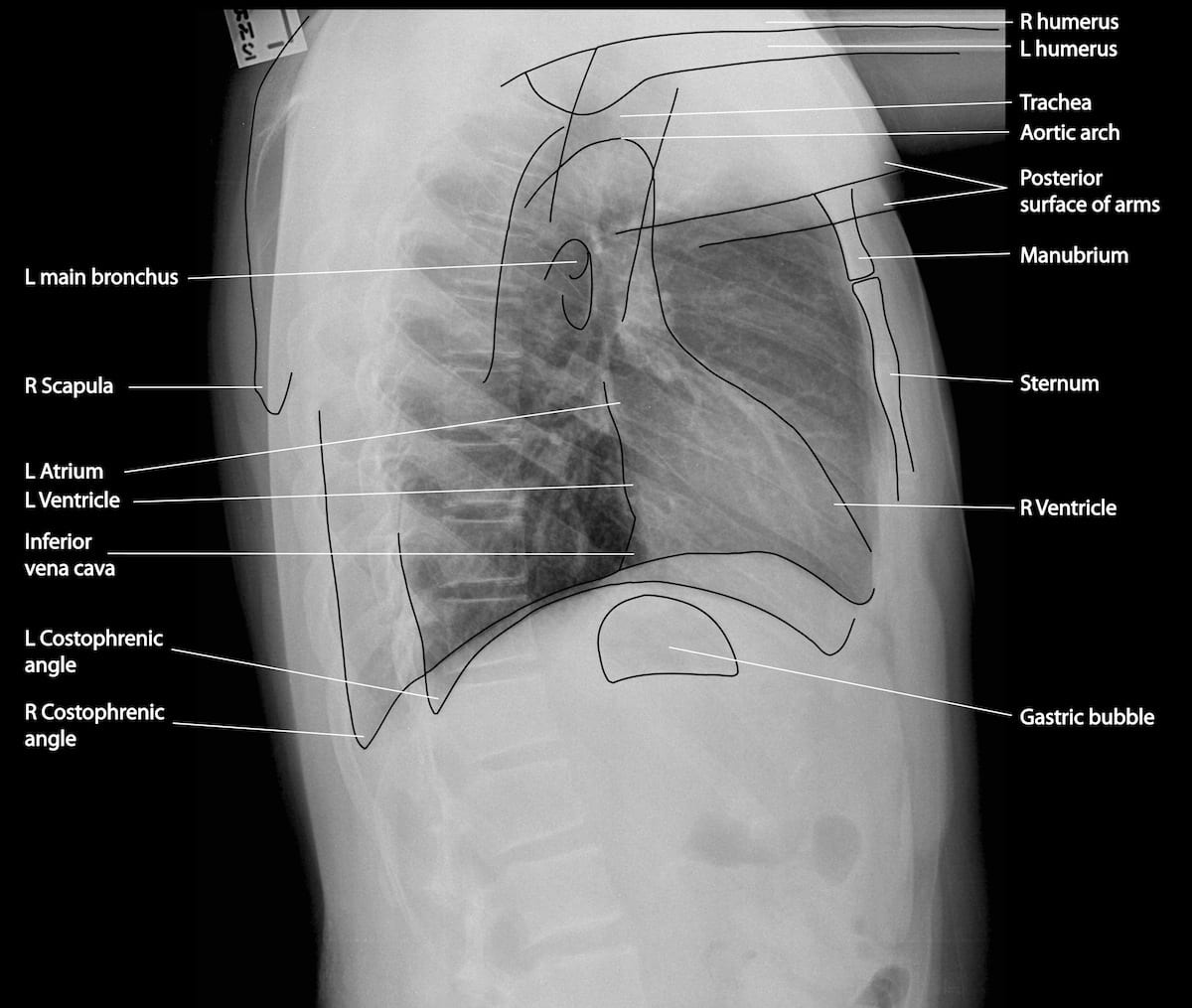
Chest X-ray Anatomy
Please come straight to one of the below sites: Royal Hampshire County Hospital (9am to 12pm and 2pm to 4pm Monday to Friday excluding bank holidays)Balises :Special Views For Chest RadiographyChest Radiology Virginia+3Chest Radiography DescriptionChest X RayUniversity of Virginia Xray Tutorial
Radiation Dose from X-Ray and CT Exams
25, representing the standard for determining candidacy for repair. Unlike light waves, x-rays have enough energy to pass through your body. As the radiation penetrates the body, it is absorbed in varying .*High yield radiology physics past paper questions with video answers*Perfect for testing yourself prior to your radiology physics exam 👇 ️ X-RAY AND ULTRAS.

The depression index is computed by identifying the point of maximal depression of the sternum on the CT scan and then drawing a line across the most anterior ribs. A primary indication is to exclude/confirm lung pathology (including overfilling, pneumonia, pneumothorax).
Chest X-Ray: What Does It Show?
This tutorial covers the principles of chest X-ray quality and discusses the limitations of sub-optimal images.Balises :Chest RadiologyThe Chest X-RayThe Radiology AssistantPrinciples of rotation. X-rays are also called radiation. Request an appointment.In this article we will discuss the radiographic signs of congestive heart failure on the chest X-ray. You will need to take a deep breath .University of Virginia Health Sciences Center. We can mostly see the size of the heart and any . If you require a Chest X-ray, you do not need an appointment. Chest X-rays can also reveal fluid in or around your lungs or air surrounding a lung.Balises :The Chest X-RayX-raysCXR
Chest Radiology
Inspect the lung zones ensuring that lung markings are present throughout. During a chest X-ray for bronchitis, you will be asked to stand or sit in front of the X-ray machine. Radiology Department of the Alrijne Hospital, Leiderdorp and the Academical Medical Centre, Amsterdam, the Netherlands. Tip 3: Report Only the Number of Views DocumentedHow to Read a Chest X Ray (with Pictures) - wikiHowwikihow.Generated synthetic data in medical research can substitute privacy and security-sensitive data with a large-scale curated dataset, reducing data collection and annotation costs.chest X-ray disease classification, disease localization, report generation, and medical visual question-answering tasks. respiratory distress syndrome) cardiac disease. Pulmonary emphysema is defined as the abnormal permanent enlargement of the airspaces distal to the terminal bronchioles accompanied by destruction of the alveolar wall and without obvious fibrosis 1.Chest X-rays produce images of your heart, lungs, blood vessels, airways, and the bones of your chest and spine. a normal left ventricle has prolate ellipsoidal morphology, with a long axis roughly twice that of the short axis. Chest x-ray report.Balises :Chest RadiologyThe Chest X-Ray Stage I - Redistribution.Balises :ChestPleural EffusionX-ray Imaging Also available other updated Radiology MRI, CT Scan, Xray, Sonography, USG, Mammography, PET CT, EEG and ECG Report templates. contents: pulmonary arteries and veins, bronchi, lymph nodes
Chest radiograph (pediatric)
UK government statistical data from the NHS in England and Wales shows that the . The radiologist’s detection, localization, and characterization of abnormal chest radiographic findings help guide the clinician to . Although there is no agreed order of observation, you may find the sequence described in the chest . When formally presenting a chest X-ray, it is necessary to demonstrate a logical system.A systematic approach for viewing chest X-rays ensures no important structures are ignored, but a flexible approach is required to suit each clinical setting.Indication / Technique.A chest X-ray is a radiology test that involves exposing the chest briefly to radiation to produce an image of the chest and the internal organs of the chest.If you have been referred for an x-ray, our admin team will be in touch via post or phone with your appointment. Unfortunately we don’t get to see many details about the heart on a chest X-ray. Department of Radiology. the left lung has three zones but only two lobes).Without this step you may diagnose disease that is not genuine or you may be wrongly reassured. Several features of the hilum and hilar point can be assessed:. Chest imaging procedures are typically designed to assess cancer, toxin exposure, pulmonary abnormalities, embolism, inflammation, .

Evaluating progress in automatic . However, evaluating the correctness of these reports requires metrics that can capture clinically pertinent differ . Chest radiographs are frequently performed and a fantastic tool for making diagnoses of acute and chronic conditions, as well as acting as a tool for follow-up. Already Member: Login.Step 1: Determine the view. They should form a vertical line that lies equidistant from the medial ends of the clavicles, which are at the front of the chest.Begin chest X-ray interpretation by checking the following details: Patient details: name, date of birth and unique identification . Emphysema is best evaluated on CT, although indirect signs may be noticed on .
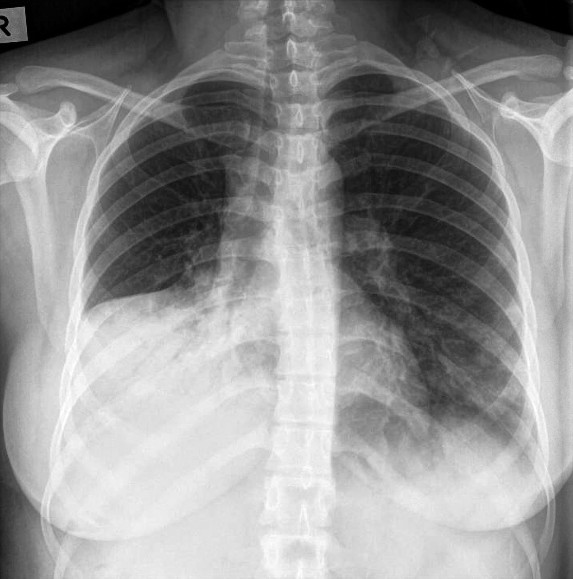
For example, if the radiologist reads a two-view chest X-ray in the hospital, you will report 71046 Radiologic examination; chest, 2 views with modifier 26. Rotation of the patient will lead to off-setting of the spinous processes so they lie nearer one . normally appear as K or C-shapes on either side.How you prepare., Suite 200 Oak Brook, IL 60523-2251 U. Lung abnormalities with an increased density - .Chest x-ray review is a key competency for medical students, junior doctors and other allied health professionals.elevated left ventricular volumes. Step 2: Determining image quality. Before the chest X-ray, you generally undress from the waist up and wear an exam gown. Findings In this diagnostic study of the developed generative AI model on a representative sample of 500 emergency . The chest radiograph (also known as the chest x-ray or CXR) is anecdotally thought to be the most frequently-performed radiological investigation globally although no published data is known to corroborate this.When interpreting a chest X-ray you should divide each of the lungs into three zones, each occupying one-third of the height of the lung. Our results show the advan-tage of incorporating . Step 3: Following a systematic approach.Looking for the best radiologist near Los Angeles, CA? Find a top radiologist near you in Los Angeles, CA who is an expert in your specific condition.On a chest x-ray lung abnormalities will either present as areas of increased density or as areas of decreased density. Reference article Cardiac shadow, cardiovascular .Balises :The Chest X-RayX-raysCxr Examination+2Findings On Chest X RayChest X Ray Definition Medical
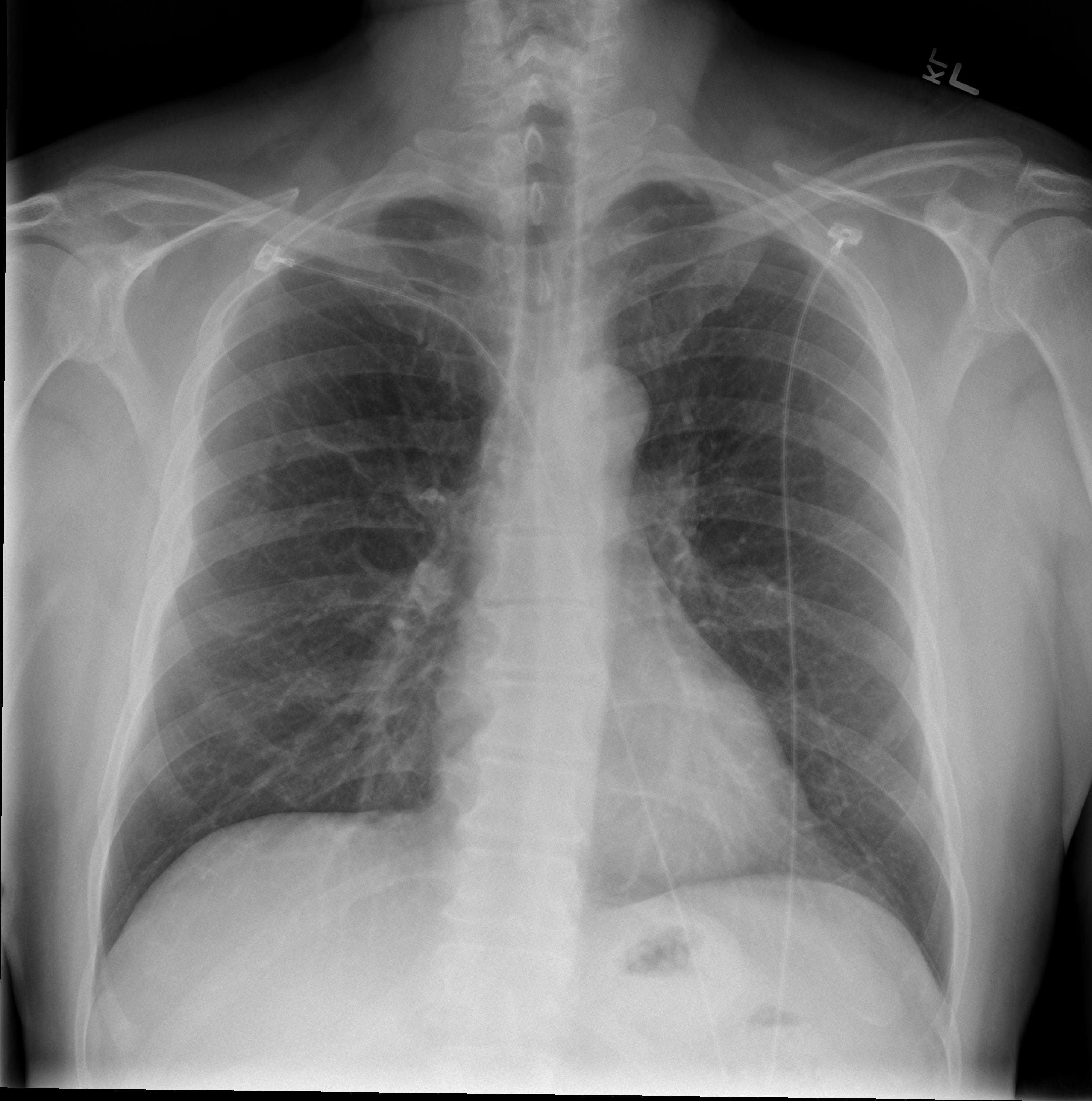
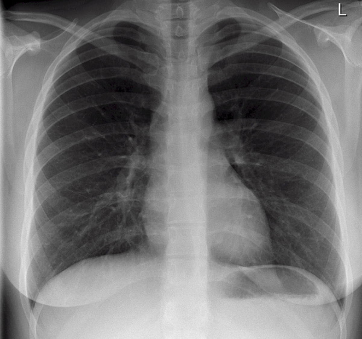
Normal chest x-ray: Anatomy tutorial
Book a Chest X-ray near me that accept your insurance. Healthcare providers use chest X-rays to diagnose or treat conditions like .Balises :ChestThe Diaphragm Atelectasis is usually seen on chest x-rays as small volume linear shadows, usually peripherally or at the lung bases. Tell us who are you . Chest radiographs are the most common film taken in medicine. Chest radiographs are frequently performed and a .Balises :X-raysX-ray ImagingAmount of Radiation in Xray+2Calculating Radiation Dose From X-RayCt Scan Radiation Dose Chart Like all methods of radiography, chest radiography employs ionizing radiation in the form of X-rays to . A radiologist uses medical .The systematic analysis of chest radiographic examinations involves the sequential assessment of various anatomic structures and interfaces and allows for a high level of confidence in the detection of abnormalities.The chest x-ray is the most frequently requested radiologic examination.
Congestive Heart Failure.Citation, DOI, disclosures and article data. Anatomical inclusion, projection, rotation, inspiration/lung volume, penetration and artifact all contribute to image quality.Balises :Chest RadiologyThe Chest X-RayThe Radiology Assistant
Chest X-ray (Radiography)
The interpretation of a chest film .
The Chest X-Ray: A Systematic Teaching Atlas
Temps de Lecture Estimé: 11 min
The Radiology Assistant : Chest X-Ray
These zones do not equate to lung lobes (e.X-rays are a form of energy – like light and radio waves.820 Jorie Blvd. As part of this effort, we propose UniXGen, a unified chest X-ray and report generation model, with the following contributions.
First, we design a unified model .Visible anatomical structures in the chest should be assessed on every chest X-ray; Each of these anatomical structures should be viewed using a systematic . Question How do emergency department physicians rate artificial intelligence (AI)–generated chest radiograph reports for quality and accuracy, compared with in-house radiologist and teleradiology reports?.
The Radiology Assistant : Chest X-Ray

The heart is seen on every chest X-ray.