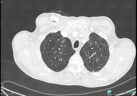
Ct scan for emphysema

Blood is taken from an artery in the wrist and can be tested to show how well your lungs are transferring oxygen into the blood and carbon dioxide out of the bloodstream. There are three emphysema types—centriacinar, panlobular, and paraseptal—that affect different parts of the lung . NJC might also prove to be a sensitive marker to detect any subtle differences that appear as emphysema progresses and could thereby become a reliable outcome predictor.
Pitfalls in Radiographic Interpretation of Emphysema Patients
Computed tomography (CT) allows direct demonstration of the presence, extent, and severity of emphysema, and CT findings correlate closely with pathologic findings. This infographic includes color coding representing the level of emphysema destruction (with darker colors representing lobes with more .
Manquant :
emphysema extra-pulmonary manifestations of COPD. As the emphysematous process progresses, the low-attenuation . comorbidities associated with COPD.The CT scan will show whether or not the airflow obstruction is due to emphysema.To determine whether visually assessed patterns of emphysema at CT might provide a simple assessment of mortality risk among cigarette smokers. Lung Procedures, Tests & Treatments.Computerized tomography (CT) scans combine X-ray images taken from many different directions to create cross-sectional views of internal organs.Low-dose CT scanning (LDCT) is an effective screening tool that decreases mortality rates from lung cancer in smokers and former smokers by revealing early-stage neoplasms .The left 3 columns (1–3) are predicted PRMs based on single inspiratory chest CT scan using deep learning, and the right 3 columns (4–6) are ground truth PRMs from real inspiratory and expiratory CT scans.
Emphysema can be identified on CT scans and shares risk factors with lung cancer, and current literature suggests that presence of emphysema may increase .
Panlobular Emphysema: Diagnosis, Treatment, and More
Also, the Fleischner framework was used to handle the training data. Panlobular (or panacinar) emphysema is a type of emphysema that affects a specific part of the lungs.Emphysema Life Expectancy | New Health Advisornewhealthadvisor.
Pulmonary Emphysema
In early stages of the . CT scan has notable potential to quantify the severity and progression of emphysema in patients. There is still debate, however, about the definition of this percentage and many researchers set it depending on the . CT scans were acquired with multidetector CT scanners . 15 Emphysema was quantified on the baseline screening CT scan as the percent low attenuation area (%LAA), defined as the percentage of lung volume with voxel density less than –950 Hounsfield units, using automated densitometry software (Imbio LLC).
Emphysema: Basics and Common Causes

Thin walled cysts with bullae larger than 1 cm (arrows) represent paraseptal emphysema and multilayered small cysts with reticulations and traction bronchiectasis (thin arrows) .Emphysema was identified in 23.The results of two studies demonstrated emphysema progression over time on CT scans and lung function decline in smokers with or without COPD (9,10).
Congenital Lobar Emphysema
Each CT scan was retrospectively visually scored by two analysts using the Fleischner Society classification system.3 million dollars per quality-adjusted life saved, 5 providing us with a sobering reminder that these ‘pretty pictures’ are .Computed Tomography Emphysema Database.Quantitative CT Analysis.Automated DL-based categorization of centrilobular emphysema from CT scans was developed.The 4 types of emphysema (causes, symptoms and . It causes permanent holes in the lower lung tissue.If you have emphysema, it’ll probably take longer. The advanced development and utilisation of quantitative CT do not simply represent regional changes in pulmonary function but aids in analysis for better patient .
Pulmonary emphysema
Emphysema is a disease of the lungs in which the air sacs in the lungs (alveoli) are permanently damaged.In the study by Oh et al ( 9 ), because 46% of smokers with emphysema and normal spirometry had only trace emphysema at baseline CT, the lack of mortality difference was not surprising. (31,32) recommended a cut-off value for emphysematous lung parenchyma on CT scans is −950 HU, which nowadays represents the standard for all different software.Congenital lobar emphysema is a rare developmental anomaly of lungs that occurs mostly due to defective bronchial cartilages. Severity of emphysema was also evaluated . 7 Alternatively, indices of lung attenuation at a given percentile along the HU histogram, such as the first percentile (P1) or 15th percentile (P15), can be used for the evaluation of emphysema.But Braga, who previously worked at the Atlantic Council think tank, stressed that OpenAI’s tools still “offered guidance on how to define intent, create .Gevenois et al.In CT imaging, the presence of emphysema is observed by a local decrease of the lung density and the diagnose is usually set as more than 5% of the lung below −950 HU, the so-called emphysema density mask. Low-dose CT scanning (LDCT) is an effective screening tool that decreases mortality rates from lung cancer in smokers and former smokers by revealing early-stage neoplasms that are amenable of . CT scans can also be used to screen for lung cancer. Early centrilobular emphysema is usually seen as small round black (low attenuating) evenly distributed holes with ill-defined borders that may appear in the central portion of the secondary pulmonary lobule around the centrilobular artery.comRecommandé pour vous en fonction de ce qui est populaire • Avis
Emphysema
The CNN was integrated into the long short-term memory (LSTM) network to predict the topic-level values from CT images.Quantitative emphysema measured on LDCT imaging of the chest can be leveraged to improve lung cancer risk prediction and help diagnose COPD in individuals . It causes a decrease in respiratory function and breathlessness. What Is a CT Scan? Computed tomography, more commonly called a cat scan or CT . For this report, we considered volumetric CT scans obtained with full inspiration (200 mAs, 120 kVp) and passive expiration (50 mAs, 120 kVp). CT settings are detailed in the COPDGene study CT protocol . Such quantification should ideally reflect the true attributes and pathologic conditions of subjects, not .How Long Does Emphysema Take to Kill You? - MDM Healthmdm-communications. This blood test measures how well your lungs are bringing oxygen into your blood and removing carbon dioxide. Emphysema, characterized by loss of lung tissue, is one of the main components of .Auteur : Nestor L.Emphysema was quantified on the baseline screening CT scan as the percent low attenuation area (%LAA), defined as the percentage of lung volume with voxel density less than –950 Hounsfield units, using automated densitometry software (Imbio LLC).Emphysema is best evaluated on CT, although indirect signs may be noticed on conventional radiography in a proportion of cases. Laboratory tests. However, progression of emphysema grade may occur over time, so an increased mortality risk after a longer follow-up time would be expected. Prior to emphysema quantification, a 3 × 3 × 3 median filter was applied to reduce .
Advances in imaging for lung emphysema
4 Recently, the cost-effectiveness ratio of lung cancer screening with CT scans was reported to be $2. It is diagnosed with a chest x-ray or ct-scan and treated with lobectomy in severe symptomatic cases. Participants
Role of Quantitative Computed Tomographic Scan Analysis in
CT scans are expensive for patients and to the healthcare system, with prices ranging from $500 to $1500 per scan.High-resolution computed tomography scan shows multiple air cysts in a mixture of paraseptal emphysema and pulmonary fibrosis in the same areas of the lungs.


Using a lower threshold than −950 HU would lead to an underestimation of the presence of emphysema, while using a higher threshold than −950 HU would lead .

A CT scan of healthy lungs looks typical in size with no inflammation, allowing the diaphragm to dome.
Subcutaneous emphysema is readily visible on CT scans, with pockets of gas seen as extremely dark low (air) attenuation areas in the subcutaneous space.
CT Scan
Materials and .Emphysema can be identified on CT scans and shares risk factors with lung cancer, and current literature suggests that presence of emphysema may increase risk of lung cancer development 6,7,8,9,10 .Consequently, the CT scan is the most sensitive and accurate option in detecting and measuring emphysema.Smoking is the main risk factor for lung cancer and COPD that includes pulmonary emphysema, chronic bronchitis, or a variable combination of both.Auteur : Katharina Martini, Thomas Frauenfelder
CT Diagnosis of Emphysema
Due to the variations in lung anatomy, manual corrections are often required to ensure successful and accurate lobe segmentation for pathological and post-treatment CT scan analysis.The imaging modalities, which can be used for the evaluation of emphysema, are manifold: Due to its high spatial resolution, computed tomography .Recently, the presence of emphysema on a CT scan has been associated with an increase in mortality in a small cohort of patients with COPD, most of whom entered the study in advanced stages of the disease.8% of patients undergoing low-dose CT and was unsuspected in 76.
Imaging of pulmonary emphysema: A pictorial review
The CT images were reconstructed in contiguous . Damage to the air sacs can't be fixed.D, Paraseptal phenotype of emphysema shows typical lesions (arrowheads) beneath pleural surface on gross specimen, and micro-CT scan shows that alveoli adjacent to lobular septa are dilated and destroyed, with sparing of center of lobule. Some individuals will . The Global Initiative for chronic obstructive lung disease (GOLD) has defined COPD as a common, preventable, and treatable disease that is characterized by persistent respiratory symptoms and airflow limitation that is due to . This activity describes the histopathology, etiology, evaluation, and management of congenital lobar emphysema . 8 As the emphysema extent . If you have advanced emphysema, your lungs will appear to be much larger than they should be.Previous studies have shown that the MLD derived from expiratory CT scans is a good predictor of pulmonary ventilation.

Computed Tomography Emphysema Database
Arterial blood gas analysis. As early as the 17th . While there is no cure for COPD, your doctor may recommend lifestyle changes, therapies, medication and/or surgery to help relieve .Lung Health & Diseases.


/ushuaia073-56d9cc195f9b5854a9caf464.JPG)