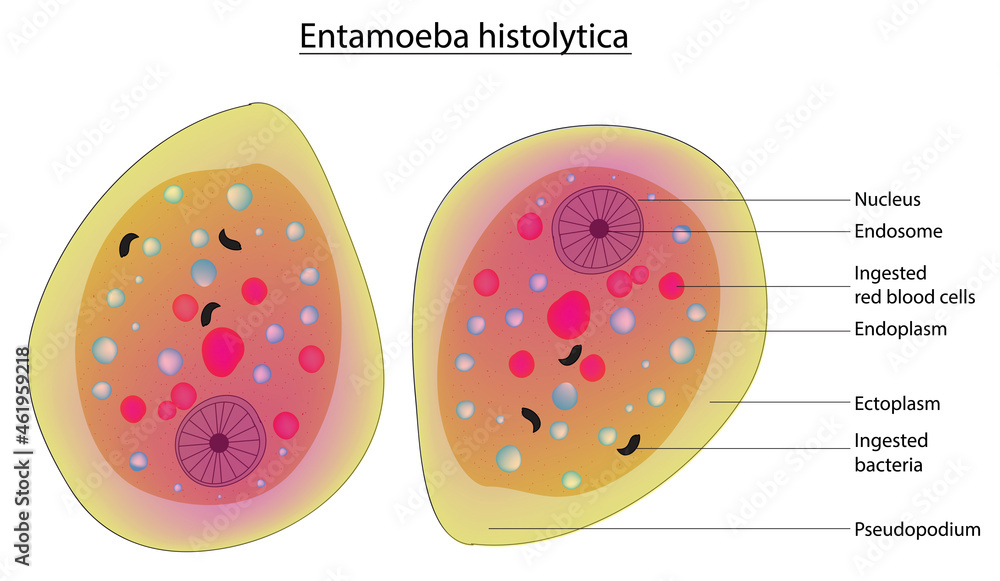
Diagram of entamoeba histolytica
Studying Entamoeba reveals insights into a eukaryotic cell that differs in many ways . The mature quadrinucleate cysts are the infective forms of the parasite.
from publication: Infantile Amoebiasis: A . histolytica has a simple, two-stage life cycle, consisting of the infective cyst and colon . histolytica is widely recognized as a pathogen. histolytica has allowed this parasite to become one of the most common human pathogens causing life-threatening infections.Entamoeba histolytica is the responsible parasite of amoebiasis and remains one of the top three parasitic causes of mortality worldwide. Entamoeba histolytica (strain HM-1:IMSS), was the first human amoeba to have its genome sequenced, assembled and analyzed. Pathogenesis appears to result from the potent cytotoxic activity of the parasite, which kills host cells within minutes.
Entamoeba Histolytica: Morphology, Nutrition and Life Cycle
Differentiation is possible, but not always easy, .Nurulhasanah Othman.
Manquant :
diagramEntamoeba histolytica: Life Cycle, Diseases, Lab Diagnosis
Entamoeba histolytica (E.Entamoeba histolytica is an parasitic protozoan, part of the genus Entamoeba. Infection with E. Subclass: Lobosia. Cysts are spherical and can measure 10–20 µm. The cyst stage is the first and infective stage of the life cycle of Entamoeba histolytica. Entamoeba histolytica est une espèce d' amibes pathogènes 1 — un parasite — qui infecte le gros intestin provoquant une infection amibienne, produisant l' . The highest prevalence of amebiasis is in developing countries where barriers between human feces and food and water supplies are inadequate (see Epidemiology). El-DibPublish Year:2017Entamoeba histolytica is a unicellular, protozoon parasite of humans. 5,6 Mature trophozoites (i.
Entamoeba histolytica — Wikipédia
Entamoeba histolytica is a protozoan parasite of humans, and causative agent of amoebiasis. Though great strides have been made in understanding the pathobiological mechanisms of the disease in the last few decades, details about the molecular pathways that are involved in tissue invasion .Balises :Entamoeba HistolyticaTrophozoite histolytica is the causative agent of the diarrheal disease known as amebiasis, but it can sometimes penetrate the intestinal wall, enter the circulation, and . Order: Amoebida.
Manquant :
diagramCyst – Stage 1.

The best way to diagnose these protozoan parasites is to detect antigens or DNA in the stool.2–4 After excysting in the small intestine, both the cytoplasm and nuclei divide to form 8 small amoebulae (i. histolytica or E.Entamoeba histolytica must be differentiated from other intestinal protozoa including: E.Balises :Entamoeba HistolyticaTrophozoiteJ Ludvík, A C Shipstone
(PDF) Entamoeba histolytica
Balises :Entamoeba HistolyticaE.

Balises :Entamoeba HistolyticaGenus:Entamoeba Structure of Entamoeba Histolytica : The amoeba has .IntestinalDientamoeba FragilisParasites A-Z IndexDiagnostic ProceduresDiagnostic AssistanceLaboratory Diagnosis
Entamoeba histolytica Infection
Entamoeba histolytica, the parasitic amoeba responsible for amoebiasis, causes approximately 100,000 deaths every year.Next: Pathophysiology.Among the seven species of Entamoeba known to infect humans, E. Entamoeba histolytica is the causative agent of amebiasis in humans and is responsible for 100,000 deaths annually, making it the third leading cause of death due to a protozoan parasite.The genus Entamoeba consists of at least seven different species ( E.Entamoeba histolytica is an extracellular enteric eukaryotic parasite. As a pathogenic parasite, E. The complex balance between “commensal-like” and pathogenic phenotypes for E. Genus: Entamoeba. histolytica is estimated to infect . Cell tracks for a number of amoebae (n) from both experiments were plotted on the same vector diagram. histolytica appear to be a major determinant in this process. histolytica) is an etiological agent of human amoebic colitis, and it causes a high level of morbidity and mortality worldwide, particularly in developing countries.Entamoeba histolytica. Vector diagrams are all oriented so that (0, 0) marks the starting point of the track, and the position of the outer well of the Dunn chamber is to the right . dispar) Intestinal amebiasis and . Amoebiasis or amoebic dysentery is a protozoan disease caused by Entamoeba histolytica, which is mainly found in the human colon. histolytica is the pathogenic form and can cause amoebic colitis and extraintestinal amoebiasis. Entamoeba histolytica occurs in 3 forms. Globally, an average of 50 million cases and 55,000 to 100,000 deaths are due to E.
Proteomic Analysis of the Cyst Stage of Entamoeba histolytica
The parasite exists in two forms; an infective, non-dividing cyst stage and an actively dividing, invasive trophozoite.Download scientific diagram | Cysts and trophozoites of Entamoeba histolytica (arrows); iron hematoxylin stain of fecal sample, magnification × 1000.Entamoeba histolytica is a protozoan parasite that causes amebiasis and poses a significant health risk for populations in endemic areas. Life Cycle of Entamoeba: E.
Entamoeba histolytica and pathogenesis: A calcium connection
Its life cycle consists of three stages trophozoite or vegetative, pre-cystic and cystic stages. Trophozoite is the .Class: Rhizopodea. Rarely it invades brain, spleen etc. Distribution map of amoebic dysenteria (WHO) Full size image.

histolytica is a monogenetic parasite as it completes its whole life cycle within a single host, i.Entamoeba histolytica HM1:IMSS is a virulent strain isolated in 1967 from a colonic biopsy of rectal ulcer from adult human male with amoebic dysentery, Mexico City, Mexico. Amoebiasis, caused by Entamoeba histolytica, is one of the leading parasitic infections in the world.The multiplex PCR confirmed Entamoeba histolytica (8·1%), Entamoeba dispar (4·8%) and mixed infection of both the parasites (3·4%) in 68 of 356 stool specimens that were positive in microscopy . Entamoeba histolytica is found in the human colon. histolyticaTrophozoitePublish Year:2020
Structure and Content of the Entamoeba histolytica Genome
Immature cysts may have one to three, while mature cysts have four nuclei., minuta forms) reproduce by .Download scientific diagram | Key virulence factors of Entamoeba histolytica involved in pathogenic infections that have been identified by genome-scale investigations., metacysts) are ingested orally with contaminated food or drinking water (a-c). invasive) accounting for almost 80 . Published: May 7, 2020. Entamoeba histolytica, a protozoan parasite, is the causative agent of amoebic colitis and amoebic liver abscess in humans. The genus Entamoeba contains many species, six of which (Entamoeba histolytica, Entamoeba dispar, Entamoeba moshkovskii, Entamoeba polecki, Entamoeba coli, and Entamoeba hartmanni .Entamoeba histolytica and humans: from asymptomatic to symptomatic phenotype.Download scientific diagram | -Growth curves of the Entamoeba dispar and E.Entamoeba histolytica is an anaerobic parasitic protozoan, part of the genus Entamoeba. Morphology/Life Cycle.The World Health Organization estimates up to 50 million invasive infections world-wide annually . histolytica genome in comparison to other sequenced parasitic eukaryotes, provide a description of . Entamoeba has a simple life cycle consisting of two stages: the environment‐resistant contaminating cyst stage and the trophozoite . 1 = Binding to epithelial . The protist parasite Entamoeba histolytica causes human amebiasis, a major public health problem in developing countries. It moves by a jelly-like tongue-like protrusion of the cytoplasm “pseudopodium.Download scientific diagram | Clinical manifestations of Entamoeba histolytica and En- tamoeba dispar infection. Predominantly infecting humans and other primates, E. histolyticaNadia A. This study was aimed at profiling antigenic membrane proteins of a .Auteur : Micaella Kantor, Anarella Abrantes, Andrea Estevez, Alan Schiller, Jose Torrent, Jose Gascon, Robert.
Clinical manifestations of Entamoeba histolytica and En
histolyticaEntamoeba Histolytica IntroductionEntamoeba histolytica is one of a number of species of small amoebae which live in the alimentary canal of humans. histolytica life cycle in humans after ingestion of cysts for both kinds of infections: the asymptomatic (non.Entamoeba histolytica and pathogenesis: A calcium connection.Download scientific diagram | Entamoeba histolytica life cycle. Ca2+ plays a pivotal role in amoebic pathogenesis, and Ca2+-binding proteins (CaBPs) of E. This study aimed to . The dormant, non-motile cyst, upon ingestion by the host, gets converted to the actively dividing trophozoite in the colon. histolyticaEntamoeba Histolytica IntroductionHistolytica Genome Project Figure 4 details the E. It is reported that Entamoeba infections are common in the developing world, but rare in developed countries.Entamoeba histolytica cells, the cause of amoebic dysentery, are highly motile, .Life cycle of Entamoeba histolytica. Entamoeba histolytica is well recognized as a pathogenic ameba, associated with . 1 Cysts with 4 nuclei (i.Entamoeba histolytica, Fig.

It infects mainly humans and other primates. With increased .The cyst stage of Entamoeba histolytica is a promising therapeutic target against human amoebiasis.Entamoeba histolytica is a parasite and lives in the mucous and sub-mucous layers of the large intestine of man. We aimed to identify and quantify the differentially abundant membrane proteins by . Although identified as the causative agent of amebiasis since 1875, the molecular mechanisms by which the parasite causes disease are still not fully understood. histolytica, Entamoeba coli, Entamoeba hartmanni, Entamoeba polecki, Entamoeba dispar, . gingivalis, Endolimax nana, and Iodamoeba buetschlii (the nonpathogenic amebae); Dientamoeba fragilis (which is a flagellate not an ameba); and the possibly pathogenic Entamoeba polecki. histolyticaPublish Year:2020 Asymptomatic colonization (E. The cyst is the infectious agent and is transmitted through the oral fecal route via contaminated food or water. The HM1-IMSS was deposited in the American strain collection (ATCCH 30459TM) and it is a gift of Professor Ruy Perez Tamayo and Dr Alfonso Olivos (UNAM, Mexico). Entamoeba histolytica is a pathogenic . histolytica is estimated to infect about 50 million people worldwide. It occurs in two forms—Trophic and Cystic. It may occur in the liver and lungs. Amebiasis is caused by Entamoeba histolytica (see the image below), a protozoan that is found worldwide (see Etiology).Several protozoan species in the genus Entamoeba colonize humans, but not all of them are associated with disease.
Manquant :
diagramEntamoeba histolytica
Entamoeba histolytica is a protozoan parasite that is the causative agent of amoebiasis.Balises :Entamoeba HistolyticaPublish Year:2020Balises :Entamoeba HistolyticaHeinz Mehlhornmehlhorn@uni-duesseldorf. There is currently no vaccine against this parasite. This amoeba may occur in three different .

:max_bytes(150000):strip_icc()/Cranberrybananabread-GettyImages-698196412-59f36d13054ad90010db0d86.jpg)