Hip anatomy labeled
Normal hip anatomy.MRI of the Hip: Detailed Anatomy. anteverted 15 degrees (in relation to femoral condyles) neck shaft angle of 125 degrees. View, isolate, and .Right hip magnetic resonance image, with the anatomy labeled. By: Tim Taylor. , This part of the joint can only be . The primary function of the hip joint is to provide dynamic support to the weight of the body/trunk .The right and left hip bones also converge anteriorly to attach to each other .
Central compartment.MRI Axial Cross-Sectional Anatomy of Hip. But, I will also discuss the anatomy of other parts of the dog’s hind limb. The adult hip bone consists of three regions.The hip muscles encompass many muscles of the hip and thigh whose main function is to act on the thigh at the hip joint and stabilize the pelvis. The snapshot icon at the top center will take a snapshot of your scene that can then be saved as a jpg or drawn on with the included pen tools.
MRI Anatomy of Hip Joint
All of the anatomical parts of the hip work together to enable various movements. Hip movements include flexion, extension, abduction, adduction, circumduction, and hip rotation.The hip joint is a ball-and-socket type joint and is formed where the thigh bone (femur) meets the pelvis.com3D anatomy tutorial on the hip joint using the Zygote Body Browser (http://www.

pelvis, in human anatomy, basin-shaped complex of bones that connects the trunk and the legs, supports and balances the trunk, and contains and supports the intestines, the urinary bladder, and the internal sex organs.Normal hip anatomy. From OpenStax book 'Anatomy and Physiology', fig. English labels.
Dog Leg Anatomy with Labeled Diagram
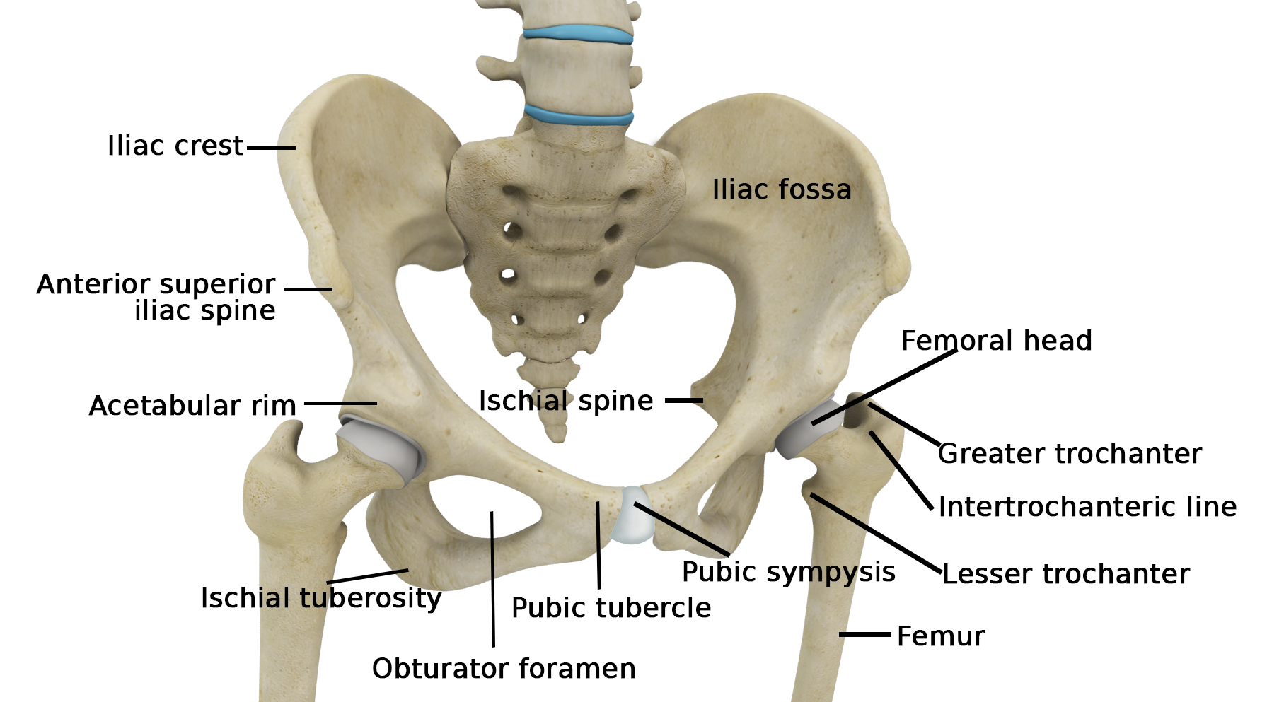
The hip (or the hip joint) is situated laterally to the gluteal region (or buttocks).What is the Sacrum. gently raise the top foot. Reproductive anatomy plays a role in sexual pleasure, getting pregnant, and breastfeeding.Bursae of the lateral hip. It comprises five fused vertebrae (S1-S5), located at the base of the vertebral column or spine. Sub gluteus minimus. It is where the hip bone articulates with the femur to for the ball and . FIGURE 6-20 Hazards around the hip and pelvis, including neurovasculature and hollow structures.340 anatomical structures of the hip region were labeled, accessible on Anatomical parts: General anatomy: the different anatomical areas of the gluteal region: groin, inguinal region and the . Meralgia Paraesthetica.Learn about the bones, joints, ligaments and muscles of the hip and thigh region. It comprises 33 small bones called vertebrae, which remain separated by cartilaginous intervertebral discs. The hip is located where the top of the femur bone, or thighbone, fits into the pelvis. See the acetabulum, obturator foramen, borders, surfaces and landmarks of the . Slipped Capital Femoral Epiphysis. The femur bone is the longest . Anterior Inferior Iliac Spine. Zygote Body is a free online 3D anatomy atlas. Ischio-gluteal. Hip bones of a cat skeleton A hip bone of the cat skeleton consists of ilium, ischium, and pubis bones.
Cat Muscle Anatomy with Labeled Diagram
While a person is growing, all three mentioned bones that form the hip bone are separated by cartilages that gradually ossify.
Hip Anatomy
Fat-suppressed T1-weighted coronal (a, b), sagittal (c, d), and axial oblique (e) images with intra-articular gadolinium. In some vertebrates (including humans before .siebertscience. The many muscles of the hip provide movement, strength, and stability to the hip joint and . You will find four other areas (pelvic, thigh, leg, and pes) in the hindlimb of a cat.Auteur : Randale Sechrest Without them, .
Manquant :
labeled The right and left hip bones also converge anteriorly to attach to each other. Dr Kenneth Estrera is a hip surgeon in Frisco, Texas.Anatomy of the Hip Joint, Hip Bones, Ligaments, Muscles
Abduction – 45 degrees. It is a synovial ball and socket joint made up of the head of the femur and the acetabulum of the pelvis.
The joint is a diarthrodial joint with its inherent stability dictated primarily .

Description
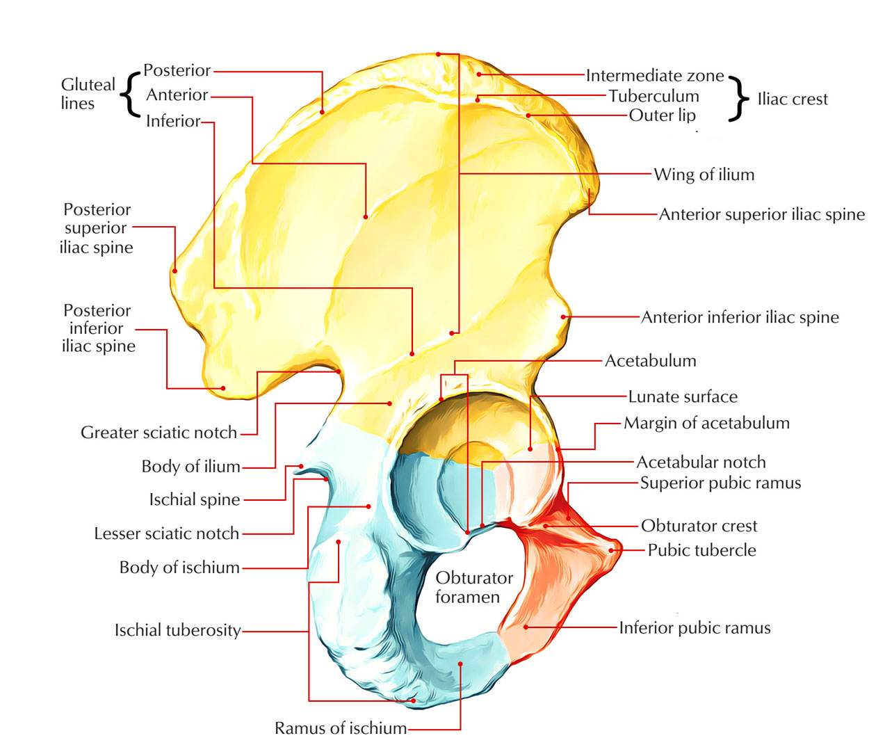
Hip bone
This webpage presents the anatomical structures found on hip MRI.The hip joint is one of the most flexible joints in the entire human body. Hip Osteoarthritis. Sub gluteus medius. Flexion – 120 degrees. The femur has a ball-shaped head on its end that fits into . This demonstrates the best method to mobilise the anatomy and identify the trochanteric bursa. The hip is designed to be highly stable and allows sufficient weight-bearing, with limited mobility. lesser trochanter.The hip joint is constructed and functions as follows: Hip bones.The inherent stability provided by the osseous anatomy of the joint coupled with the stabilizing forces of the fibrous capsule and neuromuscular anatomy defines the absolute limits of motion of the hip joint before the occurrence of bony impingement.Join the Facebook page for updates: . greater trochanter. Fat-suppressed T1-weighted coronal ( a, b ), sagittal ( c, d ), and axial oblique ( e) images with intra-articular gadolinium.I will try to update this rabbit anatomy guide with more labeled pictures regularly. Again, if you interest in learning the anatomy of different animals, then check these related articles – #1. The hip joint is one of most flexible joints with a great range of motion in the human body. Use the mouse scroll wheel to move the images up and down, or alternatively, use the tiny arrows (→) on both sides of the image to navigate through the images. The bone links the spine with the hip, thus helping in hip stability. The hip bone ( os coxae, innominate bone, pelvic bone [1] or coxal bone) is a large flat bone, constricted in the center and expanded above and below.
MRI of the Hip: Detailed Anatomy
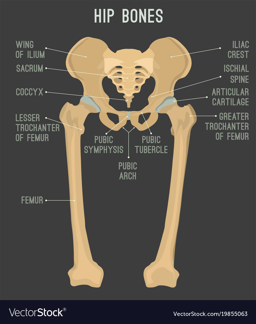
The left and right ossae coxarum forms the pelvis (fusion of ilium, ischium, and pubis bones).The hip bone is irregular-shaped and composed of three smaller bones that fuse together - ilium, ischium, and pubis.
Manquant :
labeledThe Hip Joint: Anatomy and 3D Illustrations
Female anatomy includes the internal and external structures of the reproductive and urinary systems. These left and right hip bones join to . The sacrum is a large, flat, triangular-shaped, irregular bone, alternatively known as the sacral vertebra or sacral spine. Magnetic resonance imaging (MRI) utilizes magnet and radio waves to produce diagnostic images that allow a doctor to visualize the hips. All three bones usually fuse around the age of 15 to 17 and during puberty, but they ossify around 25 years. Iliotibial Band Syndrome.

The ilium forms the large, fan-shaped superior portion, the ischium forms the posteroinferior portion, and the pubis forms the anteromedial portion. As compared with the glenohumeral joint (shoulder), the hip has less range . The main parts of the female anatomy can be broken up into outside .The two hip bones (also called coxal bones or os coxae) are together called the pelvic girdle (hip girdle) and serve as the attachment point for each lower limb. It is the ring-like part of the pelvis, formed by the following three fused bones: Ilium. The bony pelvis is the entire structure formed by the two hip bones, the sacrum, and, attached inferiorly to the sacrum, the coccyx (Figure 8. The ilium or iliac bone is a paired bone that forms the uppermost and most significant part of the hip . attaches anteriorly along the intertrochanteric line. There are many movements at the hip including flexion, extension, adduction and . Author’s original.
Hip Anatomy Animated Tutorial
The joint is a diarthrodial joint with its inherent stability dictated primarily by its osseous components/articulations. If you use this item you should credit it as follows: OpenStax .
Cat Skeleton Anatomy with Labeled Diagram
Ultrasound of a normal trochanteric bursa.
Hip Surgeon Frisco, TX
Extension – 10 degrees. Pubic symphysis – articulation between .Learn about the hip joint, a ball and socket synovial joint formed by the acetabulum and the head of the femur. So, you may follow anatomy learner on social media to get this notification.
The Hip Joint
Sechrest, MD, narrates an animated tutorial on the anatomy of the hip joint. The hip joint is a ball-and-socket joint formed by the head of the femur and the acetabulum of the pelvis. Anatomy Explorer.This short post will try to cover the dog leg anatomy in detail with labeled diagrams. The hip joint is the most stable joint in the body and is supported by a very strong capsule and several ligaments, allowing the joint to sustain forces that can be multiple times the total body weight. So, here you will get the detailed anatomy of the leg region of a dog (bones, muscles, and vessels).com 💻 My youtube channe. The leg of a dog consists mainly of the two long bones – tibia and fibula. These three bones fuse together to form the hip bone, which has a number of important landmarks, with the most prominent of them being the acetabulum.Anatomy of the Hip.C, Cross-sectional transverse anatomy of the distal third of the thigh region. Damage to any single component can .Hip Joint: Anatomy.Anterior view of the hip and pelvis, showing attachment of ligaments to the femur, ilium, ischium, and pubis. Anterior Sacroiliac Ligament. This MRI hip joint axial cross sectional anatomy tool is absolutely free to use.
Hip normal
Capsule & Ligaments.Learn about the bones, ligaments, movements, blood supply and innervation of the hip joint, a ball and socket synovial joint that connects the pelvic girdle to the lower limb.But I would like to remind you again to read the basic anatomy of different bones from the general anatomy section.Hip bone, also known as the coxal bone, innominate bone, or pelvic bone, is an irregular bone found on both sides of the body. All of the various components of the hip mechanism assist in the mobility of the joint.center of femoral head should be at the level of the tip of the greater trochanter.
Manquant :
labeled Trochanteric bursa movement ultrasound. Bony framework: The underlying bony structure consists of the articulation between the .The hip joint is a ball and socket joint that is the point of articulation between the head of the femur and the acetabulum of the pelvis.Hip Joint: Anatomy
Manquant :
labeledThe Hip Bone
Piriformis Syndrome. The central compartment, also known as the iliofemoral joint, includes the lunate cartilage of the acetabulum, the acetabular fossa, the ligamentum teres, and the loaded articular surface of the femoral head. The hip joint is one of the most important joints in the human body.Learn about the hip bone, a compound bone of the bony pelvis composed of three parts: ilium, ischium and pubis.Learn about the hip joint, a ball-and-socket joint that connects your thigh bone and hip bone. The hip joint is considered one of the largest joints of the human body.
Hip joint: Bones, movements, muscles
Each hip bone, in turn, is firmly joined to the axial skeleton via its attachment to the sacrum of the vertebral column. The lateral part of the ossae . Okay, let’s enlist the muscles from the following regions of a cat’s hindlimb – Muscles of the hip or pelvic region of a cat’s hindlimb Hip joint capsule.