Hip dysplasia adult radiology

1 Developmental dysplasia of the hip. Adult hip dysplasia and osteoarthritis.govRadiographic Parameters of Adult Hip Dysplasia - SAGE .
Dornoosh Zonoobi , Abhilash Hareendranathan , Emanuel Mostofi , .Developmental dysplasia of the hip (DDH), or in older texts congenital dislocation of the hip (CDH) , denotes aberrant development of the hip joint and results from an abnormal relationship of the femoral . Because of their low cost and ready accessibility, radiographs are the primary imaging modality used to evaluate patients with hip pain and to establish the diagnosis of hip dysplasia.Development dysplasia of the hip (DDH) encompasses a broad spectrum of abnormal anatomical development of the femoral head and the acetabulum of the hip joint.1007/s12178-016-9369-0. Acetabular deficiency and malalignment result in an abnormal force transfer across the hip joint leading to pathologically elevated cartilage stresses [ 1 , 2 ].Adult hip dysplasia and DDH have pathoanatomic similarities, but the link between them, if there is one, is not clearly understood.Winner of the Society of Skeletal Radiology Excellence Award for an Exhibit: Diagnostic Evaluation of Hip Dysplasia in the Young Adult: Emphasis on Cross-Sec - tional Imaging at the 2011 Society of Skeletal Radiology annual meeting.Hip dysplasia is a lack of femoral head coverage and disruption of hip and acetabular alignment and congruency, with severity ranging from mild subluxation in nascent at-risk hips to complete dislocation. In hip joint OA, congenital or developmental malformation is believed to constitute an individual risk factor for premature degeneration.Background and purpose — Hip dysplasia in adults is a deformity in which the acetabulum inadequately covers the femoral head. The acetabular index, also known as the acetabular roof angle or Tönnis angle , is a radiographic measurement of acetabular inclination.Hip dysplasia is a not uncommon feature in adults and can vary from subtle acetabular dysplasia to complex sequelae of developmental dysplasia of the hip. Presentation of hip dysplasia in neuromuscular conditions can be sub-clinical or associated with a limp with or without .The radiology of hip dysplasia. It is also known as congenital hip dysplasia, but . An increased acetabular angle defining acetabular dysplasia varies between authors and is reported at >42º 6 or ≥45º 7.Familiarity with the radiographic and cross-sectional imaging findings of mild hip dysplasia in the young adult may allow a timely diagnosis and implementation of treatment strategies, which may .
The purpose of this article is to review developmental dysplasia of the hip (DDH), a well-described entity previously evaluated with a standard multimodality imag- ing algorithm, .Unlike congenital dislocation of the hip, developmental dysplasia of the hip is not confined to congenital malformations and includes perturbations in development 12.Imaging parameters used in the workup for hip dysplasia and BHD include the lateral center-edge angle, To¨nnis angle, iliofemoral line, and presence of an upsloping lateral . From a previous randomized trial (n = 12,014; 1988–90) evaluating the role of hip . It also can hurt the soft cartilage, called the labrum, that rims the socket portion of the hip joint. Cross-sectional imaging, including CT and MRI, afford improved detection and characterization by providing morphologic information about . Nine out of ten cases of hip dysplasia are diagnosed during adolescence or adulthood.Despite newborn screening programs and improvements in early diagnostic imaging, there is an estimated 0.
Adult Dysplasia of the Hip
Infants are usually treated with a soft brace, such as a Pavlik harness, that holds the ball portion of the joint firmly in its socket for several months.
The Bernese periacetabular osteotomy for treatment of adult hip dysplasia
Mild hip dysplasia might not start causing symptoms until a person is a teenager or young adult. The radiological and epidemiological studies had several aims: To critically . Pediatric Imaging • Review This article is available for .Hip Dysplasia is usually diagnosed by an experienced physician using the combination of symptoms, hip examination, and x-ray findings. DDH describes a spectrum of abnormalities that involve an abnormal relationship of the femoral head to the acetabulum, which includes mild instability, subluxation, and frank dislocation. Materials and Methods In this retrospective multi-institutional study, 3D US was added to conventional two-dimensional (2D) US of 1728 infants (mean . Developmental dysplasia of the hip (DDH) occurs due to an abnormal hip development, which presents in infancy or early childhood with a spectrum ranging from dysplasia to dislocation of . Starr (vanessaleestarr@gmail. Several indices and ratios have been developed to characterize 2-D dysplastic morphology. The prevalence is sparingly . 2016; 9 (4):427–434. Doctors are more and more aware of this common condition that often escapes detection during childhood. This angle may be too high or too low in people with hip problems.1 Radiographs . The purpose of this narrative review was to detail various established criteria and parameters on anteroposterior pelvis plain . One study showed that adult patients . The frontal projection of the pelvis perm .
Developmental Dysplasia of the Hip
Hip Dysplasia: What’s With All the Angles?
This helps the socket mold to the shape of the ball.

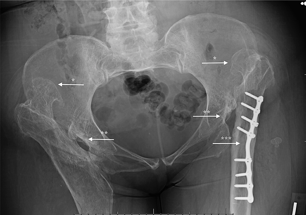
The case presented is a 53-year old woman who complained of persistent pain in the hip . Hip dysplasia in the young adult caused by residual childhood and adolescent-onset dysplasia.2% for adult hip dysplasia in our cohort and only 7% of the identified cases were described in the radiology reports.The radiographic diagnosis of mild hip dysplasia in the young adult may be subtle and is primarily based on the detection of deficient coverage of the femoral head by the acetabulum.Imaging of developmental dysplasia of the hip: ultrasound, radiography and magnetic resonance imaging.The purpose of this article is to review developmental dysplasia of the hip (DDH), a well-described entity previously evaluated with a standard multimodality . In older children and young adults, surgery may be .Dysplasia of the Hip With the Role of MRI Vanessa Starr1 OBJECTIVE.Radiographic measurements of dysplastic adult hips - .This, in combination with dysplasia, may be a source of hip pain in the young adult. It is most useful in .The normal range is 33º to 38º 6,8.The management of adult hip dysplasia is to preserve the hip and reduce pain through surgical intervention.Hip dysplasia is often diagnosed with an anterior-posterior pelvic radiograph. A CEA less than 20°, as measured on an anteroposterior radiograph of the pelvis, can be utilized to diagnose .evidence of developmental dysplasia of the right hip joint (DDH) with relatively small-sized right capital femoral epiphysis and shallow right acetabulum as well as superolateral . 2006 Dec;77(324):1-37.Developmental Dysplasia of the Hip. Musculoskeletal imaging. The radiological and epidemiological .
If you have hip dysplasia, you are not alone! Its the most common cause of hip arthritis before the age of 50. Radiology is a fundamental contribution in the .

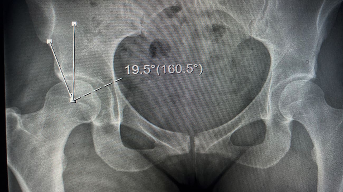
The reported incidence of this condition accounts for approximately 2–6:1000 live newborns [. Hooper N, Aroojis A, .Acetabular dysplasia in young adults occurs, despite screening for developmental hip dysplasia (DDH) in the neonatal period.of hip dysplasia, as multiple radiographic measurements are used3,34 and the adult acetabular anatomy varies according to sex and ethnicity. < 125 degrees = “coxa vara” (“varus”) There is a clear female predominance, and it usually occurs from ligamentous laxity and abnormal position in utero.20 The lateral CEA, first described by Wiberg,37 is the most com-monly utilized radiographic measure.Adult Dysplasia of the Hip is a disorder of abnormal development of the hip joint resulting in a shallow acetabulum with lack of anterior and lateral coverage.

Osteoarthritis (OA) presupposes the interaction of systemic and/or local factors.
Developmental dysplasia of the hip
Imaging parameters used in the workup for hip dysplasia and BHD include the lateral center-edge angle, Tönnis angle, iliofemoral line, and presence of an upsloping lateral sourcil or everted labrum, among many others.Developmental dysplasia of the hip (DDH) is one of the most common musculoskeletal problems in newborns.Adult hip dysplasia (AHD) results in a hip joint that has a suboptimal mechanical articulation secondary to deficient or malaligned acetabular cartilage.Radiographic Findings. [PMC free article] [Google Scholar] 11. This is called a hip labral tear.Developmental Hip Dysplasia Diagnosis at Three-dimensional US: A Multicenter Study. The literature has supported a normal lateral center edge angle (LCEA) as ≥25°, borderline dysplasia an LCEA of 18° to 25°, and an LCEA <18° as true dysplasia, though some authors will diagnose borderline dysplasia as an LCEA of 20° to 25° and true dysplasia .Citation, DOI, disclosures and article data. Hip dysplasia treatment depends on the age of the affected person and the extent of the hip damage.Abnormal Anatomical and Radiological Changes in a Rare Presentation of Adult Developmental Dysplasia of the Hip - PMC.Young adult hip disorders are increasingly diagnosed and treated as arthroscopic procedures improved. This review article describes the most useful radiographic measurements used to evaluate the adult hip.
Adults
A decreased acetabular angle . Current Reviews in Musculoskeletal Medicine. We found a prevalence of 5.
Acetabular index
When the angle is between the upper end of the normal range and an increased acetabular angle the terms borderline or indeterminate can be used 6,7.1% prevalence of hip dysplasia that goes undetected until adulthood.Familiarity with the radiographic and cross-sectional imaging findings of mild hip dysplasia in the young adult may allow a timely diagnosis and implementa-tion of treatment .International Hip Dysplasia Institute Task Force Group . Normal angle =125-135 degrees.

Hip dysplasia (HD) is such a malformation. Therefore, it is more common with oligohydramniotic . Published: 04 November 2019. Studies in radiology and clinical epidemiology Acta Orthop Suppl. Hip dysplasia can damage the cartilage lining the joint. Hence, early diagnosis provides more options as the treatment dilemma with the late presentation is very complicated with debatable prognosis. Author Steffen Jacobsen 1 Affiliation 1 Department of Orthopaedic Surgery - 333, Hvidovre University Hospital, Kettegaard Allé 30, DK-2650 . Address correspondence to V.
Hip Dysplasia
Johnson Jr DOAdult Limb Deformity & Correction
Radiographic Parameters of Adult Hip Dysplasia
Bo Yoon Ha Starr V, Ha BY 1Both authors: Department of Radiology, Santa Clara Valley Medical Center, 751 S Bascom Ave, San Jose, CA 95128.The acetabular angle, also known as the Sharp angle 6, is a radiographic measurement most commonly used when evaluating for potential developmental dysplasia of the hip (DDH) . We retrospectively compared 11 radiographic parameters describing the radiographic acetabular anatomy among hip dysplasia (26 hips undergoing periacetabular osteotomy), control hips (21 hips, requiring no rim trimming during surgical hip dislocation), hips with overcoverage (14 hips, requiring rim trimming during surgical .