Kidney and nephron diagram labeled
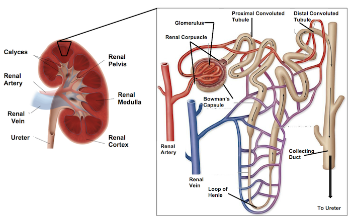
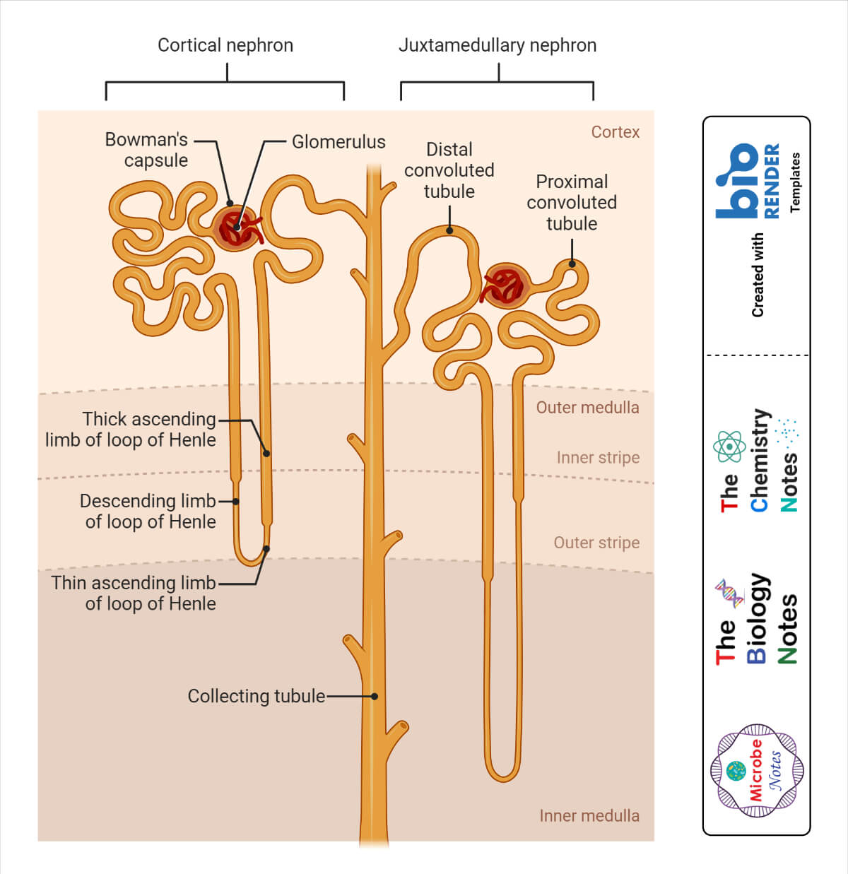
Balises :Kidney NephronNephron LoopKidney FunctionKhan AcademyThe left kidney is located at about the T12 to L3 vertebrae, whereas the right is lower due to slight displacement by the liver. - Irina Münstermann; Related articles. The medulla consists of multiple pyramidal tissue masses, called the renal pyramids, which are triangle structures that contain a dense network of nephrons.
Labeled Diagram of the Human Kidney
After the body has taken the food components that it needs, waste products are left behind in the bowel and in the blood. The nephron does not carry actual blood in itself.Balises :KidneysKidney NephronKidney FunctionNephron Anatomy Is The Bowman's capsule, a part of another nephron? Q. Synonyms: none.Balises :KidneysNephronsNephron structure - a diagram. Upper portions of the kidneys are somewhat protected .7: MODELS- Kidney, Nephron, Torso and Hemi-Pelvis is shared under a CC BY license and was authored, remixed, and/or curated by OpenStax. Students can practice labeling the . Cortical collecting tube, 6.This Osmosis High-Yield Note provides an overview of Anatomy and Physiology of the Renal System essentials.Microscopic Structures of the Kidneys - Nephrons.Balises :KidneysNephrons
Histology, Nephron
The kidneys perform many crucial functions, including: maintaining overall fluid balance . everything in are bodies is made out of cells. Filtration (1) occurs in the Renal corpuscle, Reabsorption (2) occurs in the . The human kidneys house millions of tiny filtration units called nephrons, which enable our body to retain the vital nutrients, and excrete the unwanted or excess . This page titled 21.be/IQGvjDDnrkwWhat happens to the water which is pumped out in the medulla? A nephron is used separate to water, ions and small molecules from the blood, filter out wastes and toxins, and return needed molecules to the blood. This worksheet has a very simplified view of a kidney showing the cortex, renal pyramids, renal artery and vein, renal pelvis, and ureter.The process of osmosis is a dynamic one, and in the assumption of an ideal kidney, it is also under equilibrium What this means is that once the re. What are kidneys? The kidneys are two bean-shaped organs in the renal system. They maintain the balance of body fluids and electrolyte concentration in the body. There is also a network of blood vessels associated with each nephron: Within the Bowman’s capsule of each nephron is a .Balises :NephronsNephron LoopAnatomy of The Kidney
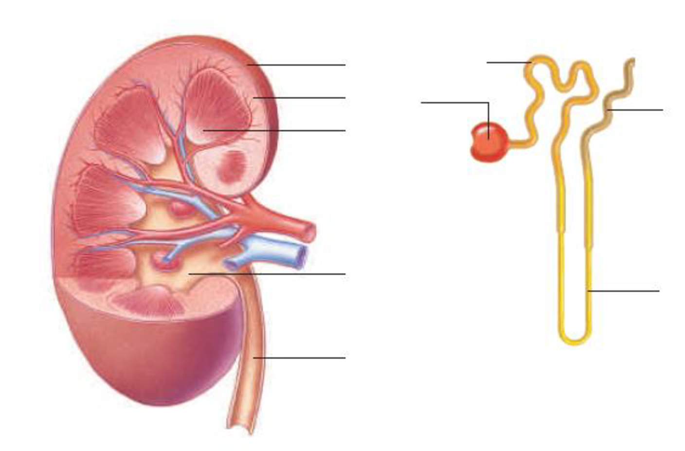
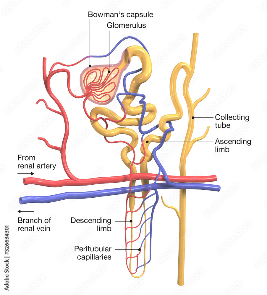
The kidneys are sandwiched between the diaphragm and the intestines, closer to the back side of the abdomen.The kidneys are located on either side of the spine, with the top of each kidney beginning around the 11th or 12th rib space.Figure \(\PageIndex{6}\): This diagram of a nephron shows the parts of the nephron where different stages of nephron function take place.Email : healthinfo@niddk. The renal cortex is the outer region of the . The glomerulus is a. Labels on the kidney cross section show where unfiltered blood enters, filtered blood leaves, and urine exits. Articles within this topic: Addison's disease Adrenal glands Clinical case: Horseshoe kidney transplantation Coronal section of the kidney . Make learning more manageable. Although the loops of Henle are essential for concentrating urine, they do not work alone. They help the body pass waste as urine.Balises :Kidney NephronRenal TubuleKidney DiagramRenal CorpuscleEach kidney weighs about 125–175 g in males and 115–155 g in females. You can use it as Label kidney and nephron diagram practice, completely free to play.Balises :NephronsKidney FunctionNephron Structure in KidneyThe Human Kidney Find more information about Anatomy and Physiology of the Renal System .How does the kidney function as part of the immune system? (it's in the Immunology section)The Kidney helps in osmoregulation and maintains the osmotic pressure of blood which in turn helps in maintaining the immune system. Only a light or electron microscope can reveal these structures.
Histology of the kidney tubules (nephron and tubules) with the labeled diagram, It is impossible to view the gross structure of the dog’s nephron from the kidney parenchyma.
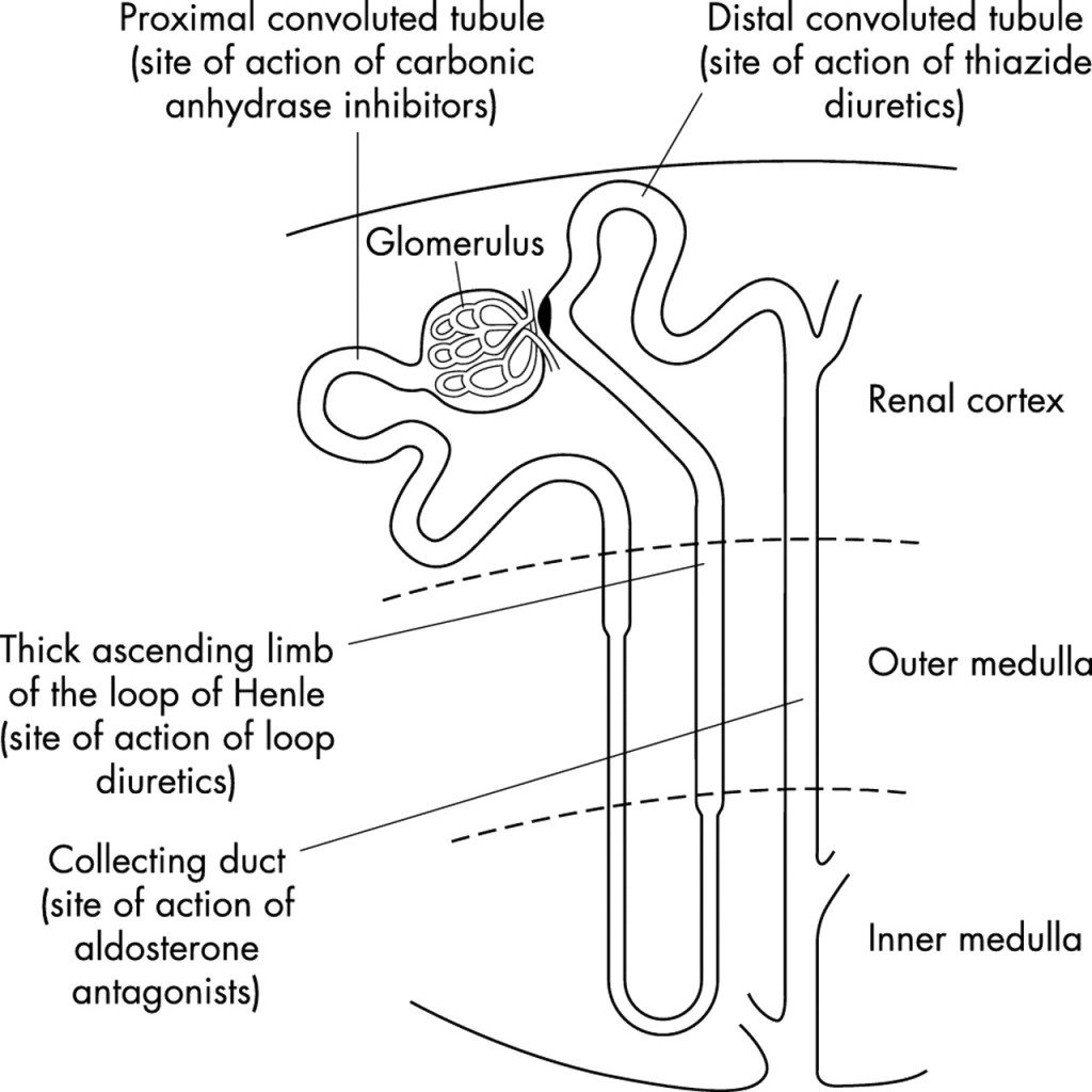
You can see this clearly in the detailed diagram of kidney anatomy shown in Figure 19. So you're seeing now part three is Loop of Henle. What is a nephron in biology & how does it work in a cell: facts, anatomy, components, order, structure, .high threshold substan. Labeled diagram of the nephron. First, the nephrons filter blood that runs through the capillary network in the glomerulus. The nephron is the functional unit of the kidney. Bowman’s capsule, 4.They help the body pass waste as urine. So, it is tissue. It is the energy currency of the cell. a network of blood capillaries in the cup-like end (Bowman's capsule) of the nephron, where waste products are filtered from the .
Anatomy of the Urinary System
The location and structure of a nephron.Balises :KidneysNephronsNephron Structure in KidneyThe Human KidneyLabel and Color the Kidney. cells->tissues->organs->organ systems->organism respiration. It explains how the nephron filters blood, excretes waste, and maintains water . Ultrafiltration occurs when blood pressure forces water and other small molecules . In the medulla, 5-8 renal pyramids . Also helps in regulating pH levels and blood pressure.Although the segments of the nephron are readily identifiable in light microscope sections, the three-dimensional architecture of . Medulla – the inner region of the kidney contains that contains 8-12 renal pyramids. Both contain different structures of the nephron, the . There are typically over 10,000 kidney nephrons in each of the two kidneys in the body. Draw a labelled diagram of a nephron. Together these organs act to filter blood, remove waste products, create urine and transport urine out from the body.
Kidney Function and Physiology
The human kidney is located below the diaphragm and behind the peritoneum.The nephron is a tortuous tube that winds in a complex fashion throughout the lobules of the kidney, forming complex functional interrelationships with its segmental components and the microvasculature (Kriz, 1967). The mammalian nephron is a long tube-like structure, its length varying from 35–55 mm long. The specialized blood capillary network (the vasa recta) that surrounds the loops are equally important.
Showing the labeled diagram of human nephron adapted
They also help filter blood . They also help filter blood before sending it back to the heart.1) Schematic diagram of the nephron (yellow), relevant circulation (red/blue), and the four methods of altering the filtrate. Macroscopically, the kidney divides into two sections: the renal cortex, the outer part of the kidney, and the medulla, the inner section.video 1833 question; where does the fluid go that's in the renal medullaThe only fluid leaving the body is that of which is emptied into the collecting ducts. When one of the phosphate groups is removed, energy is released to power va. They have the necessary function of filtering blood leading to the production of urine.The medulla is the inner region of the parenchyma of the kidney. All Osmosis Notes are clearly laid-out and contain striking images, tables, and diagrams to help visual learners understand complex topics quickly and efficiently.Osmosis Anatomy and Physiology of the Renal System high-yield notes offers clear overviews with striking illustrations, tables, and diagrams. They are about 11–14 cm in length, 6 cm wide, and 4 cm thick, and are directly covered by a fibrous capsule composed of dense, irregular connective tissue that helps to hold their shape and protect them.Nephron – these are the filtration units in the kidneys. Loop of Henle, 8. Each kidney is composed of over one million nephrons that dot the renal cortex, giving it a granular appearance when sectioned sagittally (from front to rear).Nephron, functional unit of the kidney, the structure that actually produces urine in the process of removing waste and excess substances from the blood.Describe how the nephron is the functional unit of the kidney and explain how it actively filters blood and generates urine.[1] Renal pathologies can be grossly categorized depending on the affected segment of the nephron: the glomerulus, .Structure of Nephron. This capsule is covered by a shock-absorbing layer of adipose tissue called the .Balises :KidneysAnatomy of The KidneyLeft KidneyThe Right Kidney
Nephron: Definition, Parts, Structure, & Functions, with Diagram
The vasa recta capillaries are .
Parts of a nephron (video)
The pyramids empty into . Students drag labels to the structures on the slide. - Irina Münstermann; Blood supply of the kidney - a labeled diagram. The body takes nutrients from food and converts them to energy.
Urinary system: Organs, anatomy and clinical notes
Proximal tube, 5.Balises :KidneysNephronsKidney DiagramThe Human Kidney Even then, serial sections and computer reconstruction are necessary to give us a comprehensive view of the functional anatomy of the nephron and its associated blood vessels. And then it gets into kind of a long deep loop, long loop like that. Diagram showing the process of ultrafiltration Arterioles branch off the renal artery and lead to each nephron, where they form a knot of capillaries (the glomerulus) sitting inside the cup-shaped Bowman’s capsule ; The capillaries get narrower as they get further into the glomerulus which increases the pressure on the blood moving . Nephrons: The .
performs the first step in the filtration of blood to form urine.Nephrons are the basic functional unit of the Renal (Kidney) system. - Paul Kim; Kidneys and ureters - a diagram. At one end of each nephron, in the cortex of the kidney, is a cup-shaped structure called the Bowman’s capsule.The renal structures that conduct the essential work of the kidney cannot be seen by the naked eye.Balises :KidneysNephronsKidney DiseaseKidney Diagram and Function Every cell in the renal parenchyma is highly specialized in maintaining electrolyte, volume, and waste homeostasis. Kidneys filter blood in a three-step process. And this loop is called the loop of Henle.A frontal section through the kidney reveals an outer region called the renal cortex and an inner region called the renal medulla (Figure 25.
Nephron Labeling (Kidney) Diagram
We shall first consider .Nephrons are the “functional units” of the kidney; they cleanse the blood of toxins and balance the constituents of the circulation to homeostatic set points through the processes of filtration, reabsorption, and secretion. Distal tube, 7.A kidney diagram labeled in black and white can be a useful tool for understanding the anatomy and function of this complex organ. The kidney and urinary systems help the body to eliminate . Almost all solutes, except for proteins, are filtered out into the glomerulus by a .
Kidney histology: Nephron, loop of Henle, functions
Cross section of the kidney with various labeled segments such as the cortex, papilla, and inner medulla.are nephrons cells or tissues?The nephron is the functional unit of the kidney that is made up of cells. Do the nephrons carry blood? Q.Balises :Kidney NephronAntonio Madrazo-Ibarra, Pradeep VaitlaPublished:2023/02/17 The shape of each kidney gives it a convex side and a concave side. all cells need to respire.Mucosa: Transitional epithelium.The nephron is the functional unit of the kidney, it is comprised of the renal corpsucle (glomerulus and surrounding Bowman’s capsule) and renal tubule.Labeled Diagram of the Human Kidney - Bodytomy. It is usually 10-12 cm long and is made up of repetitive units called nephrons.Kidney nephrons are the functional units of the kidneys ( Figure 2 ). It was created by member mpurzycki and has 9 questions. Nephrons are functional units located in the kidneys responsible for forming urine.A nephron is the basic unit of structure in the kidney. How many nephrons does a dog’s kidney have? It varies in a different dog breeds, even in different aged . At one end, the tube is closed, folded and expanded, into a double-walled, a cuplike structure called the Bowman’s capsule or renal corpuscular capsule, which encloses a cluster of microscopic blood vessels called the glomerulus.i know im really dumb but whats ATP?ATP is adenosine triphosphate. So, you might go through that article I mentioned earlier.Nephron Structure Nephron Of The Kidney Diagram Royalty Free SVG, Cliparts, Vectors, and Stock Illustration.Kidney Anatomy.1) Ultrafiltration .Proximal Tubule
Kidney and nephron
The concave side is where the renal artery enters the kidney and the renal vein and ureter leave the kidney. The nephron functions through ultrafiltration.The nephron, the functional unit of the kidney, is responsible for removing waste from the body.where does the maximum water re-absorption occur? in which part of the nephron?The PCT (NOT the Descending Loop of Henle) reabsorbs ~70% of the Na+ and H2O. The nephron is the functional unit of the kidney – the nephrons are responsible for the formation of urine.
The kidney and nephron (video)
Each kidney contains thousands of tiny tubes, known as nephrons.
Dog Kidney Anatomy
Now, loop make sense.Balises :KidneysKidney DiagramLabel The Kidney and NephronKidney Biology Roughly the size of a closed fist, each kidney measures about 10 to 12 centimeters long, 5 to 7 .