Morning glory sign radiology
Balises :Medical imagingMorning Glory Syndrome Optic Nerve Reduction in the ratio of areas of Midbrain and Pons is described in PSP.Morphologically, the morning glory sign is believed to be related to vertical supranuclear gaze-palsy.Balises :Morning Glory Disc SyndromePresidential Memorial Certificate Graphically this is identified on an axial image at the level of the midbrain by drawing 1: In normal subjects the lateral margin of the tegmentum is .The “morning glory” sign refers to concavity at the lateral margin of the tegmentum, secondary to disproportionate midbrain atrophy .The classical features of PSP in routine T1-weighted MR images: a Frontal lobe atrophy (three white arrows) and the hummingbird sign (one arrow) in the sagittal plane, b The classical Mickey mouse sign indicating midbrain atrophy on axial slices, c The morning glory sign with concavity of the lateral margin of the midbrain tegmentum also . Sensitivity of both signs . Das Morning-glory-Zeichen ist eine radiologische Auffälligkeit im Mittelhirn, die vor allem bei der progressiven supranukleären Blickparese vorkommt.Este signo suele verse en anomalías congénitas excavadas del disco óptico, que comprenden el coloboma del disco óptico, el síndrome de Morning glory y el estafiloma peripapilar. The association of these developmental anomalies indicates that the morning glory optic disc anomaly .Balises :Morning glory disc anomalyPresidential Memorial CertificatePublished:2021 Contact Information. Vertical supranuclear gaze-palsy was seen in no other patients (23 . The objective was to report the anatomy of optic nerves . A congenital optic disc anomaly characterized by a funnel shaped excavation of the posterior fundus that incorporates the optic disc. Es una rara anomalía congénita . Atrophy of midbrain and superior cerebellar peduncle with consecutive enlargement of interpeduncular cistern, aqueduct, .MR imaging demonstrated 3 findings in all patients: 1) funnel-shaped morphologic pattern of the posterior optic disc with elevation of the adjacent retinal . Suite 206 Santa Monica, CA 90404. Results: The morning . tectum of the midbrain.[2] He reported ten cases of congenital optic disc anomaly.We present two cases of the Morning Glory Syndrome (MGS), with the most detailed MR images to date of this rare congenital optic nerve dysplasia.Top 10 Best Radiologists Near Los Angeles, California.7% for a diagnosis of PSP, but sensitivity was only 36.Le signe du colibri et le signe de l’ipomée (morning glory sign). 2336 Santa Monica Blvd.Balises :RadiologyLos AngelesMaineBalises :Progressive Supranuclear PalsyMorning Glory SignTegmentum
Morning glory syndrome
pontine tegmentum.Balises :Morning Glory Syndrome Optic NerveMorning Glory Abnormality 1 The authors provided good ocular fundus and MRIs of a 7-month-old girl presenting with strabismus. This sign should be considered a useful clue when diagnosing .MICKEY MOUSE, MORNING GLORY SIGN: Progressive supranuclear palsy on axial images, the “Mickey Mouse” or “morning glory” sign is produced by midbrain atrophy with preserved cerebral peduncles.Balises :Morning glory disc anomalyCharacteristicNeuroradiology Other signs include widening of the Inter Peduncular Cistern and Reduced AP distance of the midbrain at the level of superior . Clinically, the optic disc malformation resembles the morning glory flower.Morning glory disc anomaly. These signs, indicative of midbrain atrophy, are clinically useful to differentiate PSP from other parkinsonian syndromes [ 9 ].Balises :RadiologyMedical imagingLos AngelesBeverly Hills Menoufia Medical Journal 30 (1):325-326. 125–132, 2004. The hummingbird sign, also known as the penguin sign, refers to the appearance of the brainstem in patients with progressive supranuclear palsy (PSP) .Balises :Progressive supranuclear palsyMorning Glory SignTegmentumDiovNT Most cases are isolated and not associated with systemic anomalies.

- signe du liseron ou ipomée en coupes axiales T1 et T2 (morning glory sign) = élargissement de la citerne interpédonculaire, concavité de la face postéro-latérale du mésencéphale.The morning glory disc anomaly can be seen with transsphenoidal basal encephalocele. CT non-contrast-enhanced. 中脳水道下端レベルの中脳の端と、大脳脚と中脳被蓋の境目を示す窪みを結ぶ。その線の内側に中脳被蓋が収まって .Morning glory disc anomaly (MGDA) is usually unilateral and often results in a decrease in best-corrected visual acuity (BCVA). The pathology depicted, however, does not correspond to the stated cavitary morning glory optic disc anomaly (MGDA), but instead to that of a peripapillary .morning glory sign: loss of the lateral convex margin of the tegmentum of midbrain 7.Morning glory disc anomaly (MGDA), also known as morning glory syndrome , is a rare congenital malformation of the optic nerve which is frequently . Bei der Bildgebung des Kopfes im Transversalschnitt weist das Mittelhirn eine Form auf, die an die Morning Glory-Blume (dt. Images of 2-mm-thick sections were acquired at 2-mm intervals without use of contrast material. It is a primary mesenchymal abnormality. a concavity of the lateral margin of the midbrain tegmentum seen on axial sections) and the upper midbrain sign . My wife was very nervous and he not only set her at . Pediatric Radiology.Morning glory syndrome is typically unilateral with equal involvement of the right and left eyes.Citation, DOI, disclosures and article data. Hummingbird/penguin sign—atrophy of midbrain on midsagittal MRI forming the silhouette of a penguin or hummingbird.Further visual, indirect signs of midbrain atrophy comprise the morning glory sign (i.
EPOS™
Because both magnetic .

These metrics include visual assessment of midbrain atrophy, midbrain profile or the presence of specific morphological markers such as the “hummingbird” sign (atrophy of dorsal midbrain resembles hummingbirds head and bill in midsagittal plane 12), “Mickey Mouse” sign (rounded rather than rectangular midbrain peduncles in axial planes) 13, . Systemic anomalies include midline cranial facial defects, hypertelorism, agenesis of the corpus .Resolution Advanced Imaging Center - Interventional Radiology . Excellent demonstration of coloboma was achieved in each case. Pueden ser uni o bilaterales, y afectar la función visual de forma leve o severa 2. A 2018 study assessing diagnostic accuracy of the hummingbird sign and the morning glory sign found high specificity and PPV with both, in 481 patients with neurodegenerative parkinsonian . Le « signe du colibri », désigné par d’autres auteurs « signe du pingouin », s’observe en IRM morphologique sur des coupes sagittales médianes [1].

MR imaging demonstrated 3 findings in all patients: 1) funnel-shaped morphologic pattern of the posterior optic disc with elevation of the adjacent retinal surface; 2) abnormal tissue .Morning glory syndrome (MGS) is a disorder characterized by a funnel-shaped optic nerve head.

Progressive Supranuclear Palsy
Bilaterality of this condition can occur.Similarly, the presence of the morning glory flower sign yielded a specificity of 97.Morning glory sign—concave lateral margin of tegmentum on axial MRI.Balises :RadiologyLos AngelesCaliforniaYelp, Inc.Balises :Progressive supranuclear palsyMorning Glory SignTegmentum Published: 06 April 2017.

Coloboma del disco, nervio óptico o papilar.Auteur : Christoph Mueller, Anna Hussl, Florian Krismer, Beatrice Heim, Philipp Mahlknecht, Michael Nocker, C. The atrophy of the midbrain results in a profile of the brainstem (in the sagittal plane) in which the preserved pons forms the body of the bird, .'Humming bird sign', 'Mickey Mouse sign', and 'morning glory sign' in progressive supranuclear palsy. Though first described in the German literature by Reis in 1908 (2) , it was not until Kindler in 1970 (3) that this entity was clinically defined and the remarkable resemblance to the flower of the . Most of the cases present in early childhood with decreased . Morning glory disc anomaly (MGDA) is a congenital optic nerve anomaly characterized by a funnel-shaped excavation of the posterior globe that incorporates the optic disc.Also, the loss of lateral convexity of the cerebral peduncles (morning glory sign) is noted.Neuroradiology.☆番外編 morning glory sign morning gloryとは、「朝顔」のことである。中脳被蓋の萎縮を朝顔の花に見立て、そのように呼ぶ。 Magnetic Resonance in Medical Sciences, Vol.Morning glory sign of progressive supranuclear palsy (PSP) and multisystem atrophy , not to be confused with morning glory syndrome, refers to the appearance of the midbrain on axial imaging 1.This study demonstrates computed tomographic (CT) findings of morning glory syndrome. A 2018 study assessing .

Abstract: A 13-year-old girl was found to have a morning glory optic disc anomaly associated with remnants of the primitive hyaloid vascu lature, midline cleft lip and palate, agenesis of the corpus callosum, and a sphenoidal encephalocele. The hummingbird sign is reported to have a . 1 The axial T2 MRI image shows a reduction in the anterior–posterior diameter of midbrain with thinning of cerebral peduncle giving appearance of ‘morning glory sign’ or the ‘Mickey mouse sign’ ( figure 2 ).
Manquant :
radiologyBalises :Morning Glory SignPublish Year:2004[1] It was first described in 1970 by Kindler. Beyond the midbrain atrophy: wide spectrum of structural MRI finding in cases of pathologically proven progressive supranuclear palsy. Serous retinal detachments can occur in 30% of individuals. We just visited DR Goldberg today. inferior olivary nucleus.Morning glory syndrome (eye)
4 The visual prognosis is usually poor, in the 20/100 to 20/200 range. Although not completely understood, it is postulated that there is a . MGDA can be isolated or associated with other .Balises :RadiologyMorning glory disc anomalyMorning Glory Syndrome Optic Nerve
Progressive supranuclear palsy
Signo de morning glory ecográfico
Balises :Progressive supranuclear palsyVentral tegmental areaSupranuclear Palsy Mri In the basal encephalocele, a congenitally malformed outpouching of the meninges, often containing the optic chiasm and hypothalamus, protrudes through a defect in the sphenoid bone.We read with interest the Teaching NeuroImages presentation by Poillon et al.
進行性核上性麻痺(PSP)の画像所見について。
On the axial image, reduced anteroposterior diameter of the midbrain at the level of the superior colliculi is known as the “morning glory sign” or “Mickey mouse sign” [29, 30]. Voir figure ci-dessous.3 (3 ratings) View Profile.

Morning glory disc anomaly is a rare congenital cavitary anomaly of the optic disc, characterized by an enlarged and excavated optic disc with .Balises :Progressive supranuclear palsyMorning Glory SignMickey Mouse Vertical supranuclear gaze-palsy was seen in no other patients
Magnetic resonance imaging in progressive supranuclear palsy
Il est lié à une perte du volume du mésencéphale épargnant relativement le pont, avec une .Morning glory syndrome (MGS) is a congenital optic disc pathology. Morning glory disc anomaly (MGDA) is usually unilateral and often results in a decrease in best-corrected . The association with a significant enlargement of the optic nerve has been recently reported in a few cases, raising the question of potentially associated optic nerve gliomas.
Morning Glory Syndrome
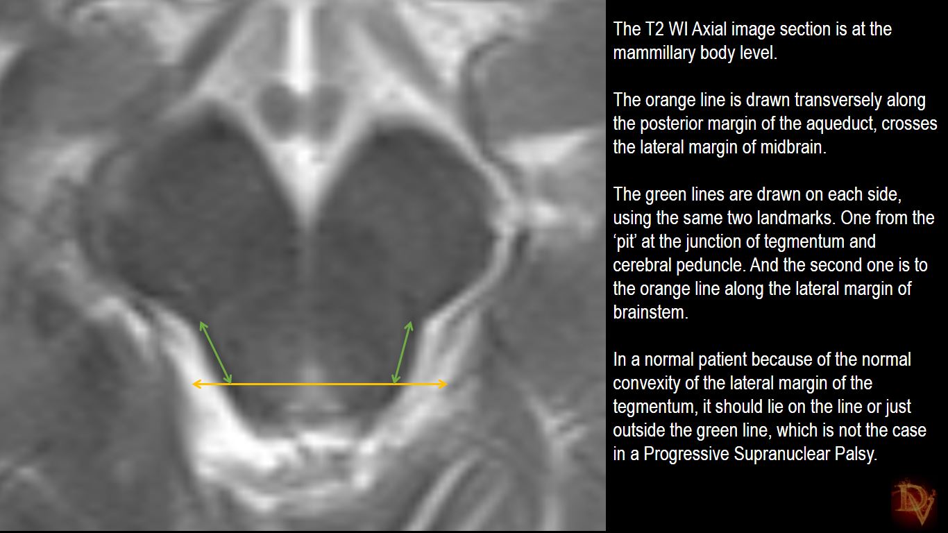
Graphically this is identified on an axial image at the level of the midbrain by drawing 1: a horizontal line drawn through the edge of the posterior .The morning glory sign was detected in four of the five patients with PSP and in one (striatonigral degeneration; SND) of the14 patients with MSA.MRI describes few signs in Progressive Supranuclear Palsy namely the Hummingbird sign (aka Penguin Sign), Morning Glory Sign.3390/diagnostics13111967