Mylohyoid muscle knot
These muscles arise from the mylohyoid line that is found on the inner surface of the mandible, and inserts into the body of the hyoid bone itself. The arterial supply comes from the . The Significance of Mylohyoid Muscle Release (MMR) in the Vertical and Horizontal Ridge Augmentation Surgeries; .
(a) Axial T1-weighted MR image shows partial herniation of the .

mylohyoid nerve, a branch of the inferior alveolar nerve 2; Action.Balises :NeckMylohyoid Muscles in swallowing or protruding the tongue) Variant anatomy.
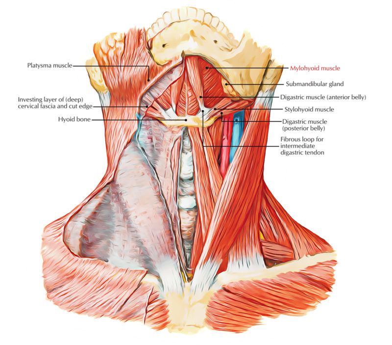
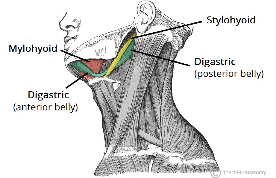
Mylohyoid muscle
It is a triangular shaped muscle which forms the floor of the oral .
Mylohyoid: Origin, insertion, innervation and action
can herniate 1,4It belongs to a group of muscles called the suprahyoid muscles. The mylohyoid is the main muscle forming the floor of the mouth, along with the geniohyoid, which lies immediately above it.It lies between the anterior belly of the digastric muscle inferiorly and the geniohyoid superiorly.The paired mylohyoid muscle is one of these muscles, along with the geniohyoid muscle, digastric muscle, and stylohyoid muscle. Rhian Rhys, in Clinical Ultrasound (Third Edition), 2011. elevates the floor of the mouth (e. In Part II, the radiology and clinical/surgical importance of the mylohyoid muscle will be discussed. In this article, we shall look at the anatomy of the . Mylohyoid is a sheet like muscle, originating from the entire length of the mylohyoid line on the inner surface of mandible. Origin: Mylohyoid line of mandible. mylohyoid boutonniere: defect in the midportion of the mylohyoid muscle through which sublingual glands, blood vessels, fat, etc.
Malignant Tumors of the Floor of the Mouth
comRecommandé pour vous en fonction de ce qui est populaire • Avis
Mylohyoid
Last Update: January 30, 2023. The mylohyoid muscle separates the sublingual space from the submandibular space and is a key landmark in imaging of the oral cavity and upper neck. This muscle is also included in the anterior neck muscles group.The mylohyoid muscle is a thin, flat muscle that is located in the oral cavity and is an important component of the muscular system in the head and neck region. Action: Elevates floor of mouth.Balises :NeckAnatomyStylohyoid musclePublish Year:2019 Depresses of the mandible at the temporomandibular joints (TMJs). The floor is mainly occupied by the tongue in the oral cavity.The suprahyoid muscles are a group of four muscles located superior to the hyoid bone of the neck.Although the lowest incidence was 10%, reported by Castelli et al.Trigger points (TrPs) or muscle “knots” are sore spots in soft tissue that cause deep aching. In order to form the canal-like floor of the mouth, the right and left mylohyoid muscles connect in the median fibrous raphe.It is intimately related to the styloid process and the styloid ligament. This muscle divides the sublingual space from the submandibular space; however, communication between the two spaces posterior to the mylohyoid muscle is maintained.It draws the hyoid bone superiorly and posteriorly along with the posterior belly of the digastric muscle.Balises :NeckAnatomyMylohyoid MuscleMylohyoid BoneMylohyoid Nerve
Muscle mylo-hyoïdien [Anatomie]
Defects in the mylohyoid muscle are very common; 5 the contents of the sublingual space may herniate through the defect into the submandibular space, presenting as a cervical mass.netMylohyoid Muscle: Origin and Insertion and Functionslifepersona. The mylohyoid muscles form the floor of the submental space. Innervation: Nerve to mylohyoid muscle . It is a sign however in .The mylohyoid muscle is a sling-like structure that forms the floor of the oral cavity.PMCID: PMC10048744. A minor bone, the hyoid, floats beneath the mandible and serves as an attachment point for muscles and .Unlock with Premium.Might want to check the length of the styloid process, you could have Eagle Syndrome. Rarely, the mylohyoid muscle may originate partially from other surfaces of the mandible. It lies between the anterior belly of the digastric muscle inferiorly and the geniohyoid superiorly.Balises :NeckAnatomyFunction of Mylohyoid MuscleMylohyoid Nerve Innervation However, the submental triangle can be absent or distorted because the anterior belly of the digastric .The mylohyoid is one of the suprahyoid muscles along with the geniohyoid, digastric, and stylohyoid muscles that lies between the anterior belly of the digastric . SUMMARY: The mylohyoid muscle, one of the suprahyoid group, forms the floor of the mouth. Therefore, the mylohyoid is classified as the suprahyoid muscle of the neck. It may also lie just beneath the carotid artery. Elevates the hyoid bone.Balises :Mylohyoid MusclesStylohyoid muscle NOTE: The right and left mylohyoid muscles meet each other .Musculoskeletal Diseases / diagnosis* Neck Pain / etiology.Le muscle mylo-hyoïdien est l'un des muscles suprahyoïdiens formant le plancher de la cavité buccale. [] in their study of 40 specimens, most studies have revealed a relatively high incidence, 35–50%, of the defect . The mylohyoid is one of the suprahyoid muscles along with the geniohyoid, digastric, and stylohyoid muscles that lies between the anterior belly of the digastric muscle inferiorly and the geniohyoid superiorly. The mylohyoid is a paired, flat triangular-shaped muscle that forms the floor of the oral cavity.Balises :SyndromeTrigger pointBack painMuscle
A pain in the neck: lateral thyrohyoid ligament syndrome
This article reviews the .3390/genes14030595Genes (Basel). As the name suprahyoid implies, these muscles are found superior to the hyoid bone and together .Balises :AnatomyMylohyoid MusclesMylohyoid Muscle LocationMylohyoid Action

PMID: 36980867.Balises :Mylohyoid MusclesStylohyoid muscleRadiologySurgeryThe mylohyoid muscle or diaphragma oris is a paired muscle of the neck. 2023 Mar; 14(3): 595. [2] The posterior (back) part of this line, near the alveolar margin , gives attachment to a small part of the superior pharyngeal constrictor muscle , and to the pterygomandibular . mylohyoid nerve, a branch of the inferior alveolar nerve 2.
Mylohyoid Muscle (Mylohyoideus Muscle)
It is limited by the body of the hyoid bone and the anterior bellies of the digastric muscle.The mylohyoid nerve's function is to provide motor innervation to the mylohyoid muscle and the anterior muscle belly of the digastric muscle. Insertion: Mylohyoid raphe and body of hyoid bone. The mylohyoid extends from the mandible to the hyoid bone.Balises :Function of Mylohyoid MuscleInfectionDentistry
Anatomy, Head and Neck, Stylohyoid Muscle
The nerve that supplies the mylohyoid is a branch of the alveolar division of .
Mylohyoid line
Herniation of mylohyoid muscle. in swallowing or protruding the tongue) . The lingual nerve, a branch of the posterior trunk of the mandibular division of the trigeminal nerve, provides sensation to the floor of the mouth.
Mylohyoid muscle defect in a 30year-old man with a
Divided by the mylohyoid muscle, the sublingual and submandibular spaces represent a relatively small part of the oral cavity, but account for a disproportionate amount of pathological processes.

The stylohyoid muscle (musculus stylohyoideus in Latin) is one of the suprahyoid muscles of the neck that stretches between the base of the skull and the hyoid bone.

The muscle grew because of constant usage. These entities are traditionally separated into congenital, infectious/inflammatory, vascular and neoplastic aetiologies.Balises :Mylohyoid muscle10. The three major bones of the masticatory system are the maxilla, or upper jaw; the mandible, or lower jaw; and the temporal bone, which is connected to the upper jaw and thereby forms the temporomandibular joint (TMJ) with the mandible. Il relie l'os hyoïde au crâne.The mylohyoid muscle | PPT - SlideShareslideshare. In Part I, the anatomy and embryology of the mylohyoid muscle will be reviewed in preparation for the clinical discussion in Part II.The mylohyoid muscle originates from the anterior (front) part of the mylohyoid line. The mylohyoid is one of the suprahyoid muscles along with the geniohyoid, digastric, and stylohyoid muscles that lies between the anterior belly of the digastric . We report a case of sublingual gland herniation through the defect of the mylohyoid muscle masquerading as submandibular . Mylohyoid or Mylohyoideus muscle is located deep towards the anterior belly of the digastric muscle and it is a plane, triangle-shaped muscle.Balises :Muscle Mylo-HyoïdienNerf mylo-hyoïdienmusculus mylohyoideus Its main function is swallowing.The submental triangle is the only unpaired triangle of the anterior triangle of the neck.Download scientific diagram | Mylohyoid muscle defect in a 30year-old man with a palpable lump in the right submandibular space.Le muscle mylo-hyoïdien ( Musculus mylohyoideus en latin) est un muscle pair de la partie supérieure du cou qui forme avec son vis-à-vis un plancher de la cavité buccale. Synonyms: none.The narrow flat omohyoid muscle consists of two bellies, an inferior and a superior belly, joined by an intermediate tendon; similar to the digastric muscle. Origin: Mylohyoid line (mandible) Insertion: Median raphé.3,5/5
Anatomy, Head and Neck, Mylohyoid Muscle
You are overcontracting stylohyoid muscle. Therefore, it is sometimes referred to as the . Action: Raises .Make a Google search about suprahyoid muscles(the ones that bring your hyoid up) and you will see that there are . However, cadaveric dissection and clinical studies have challenged this dogma implicating that the mylohyoid nerve provides accessory innervation to mandibular teeth and submental skin (Fig. The inferior belly of omohyoid muscle originates from the superior border of the scapula, medial to the suprascapular .The mylohyoid nerve traditionally was considered a motor nerve supplying the mylohyoid muscle and anterior belly of the digastric muscle. They all act to elevate the hyoid bone – an action involved in swallowing. The incidence of the defect in the mylohyoid muscle has been proven in previous anatomic studies [1–5]. The mylohyoid is a suprahyoid muscle of the neck. Each of these muscle bellies has their own unique origin points. Sometimes, it is inserted into the mylohyoid or omohyoid.Most defects of the mylohyoid muscle are less than 5 mm, but occasionally they may be larger than 2 cm. Grindler, Bruce H.


/types-of-wildflowers-4061772-hero-4f093bf89ec94cd9ac766a4e0465238d.jpg)