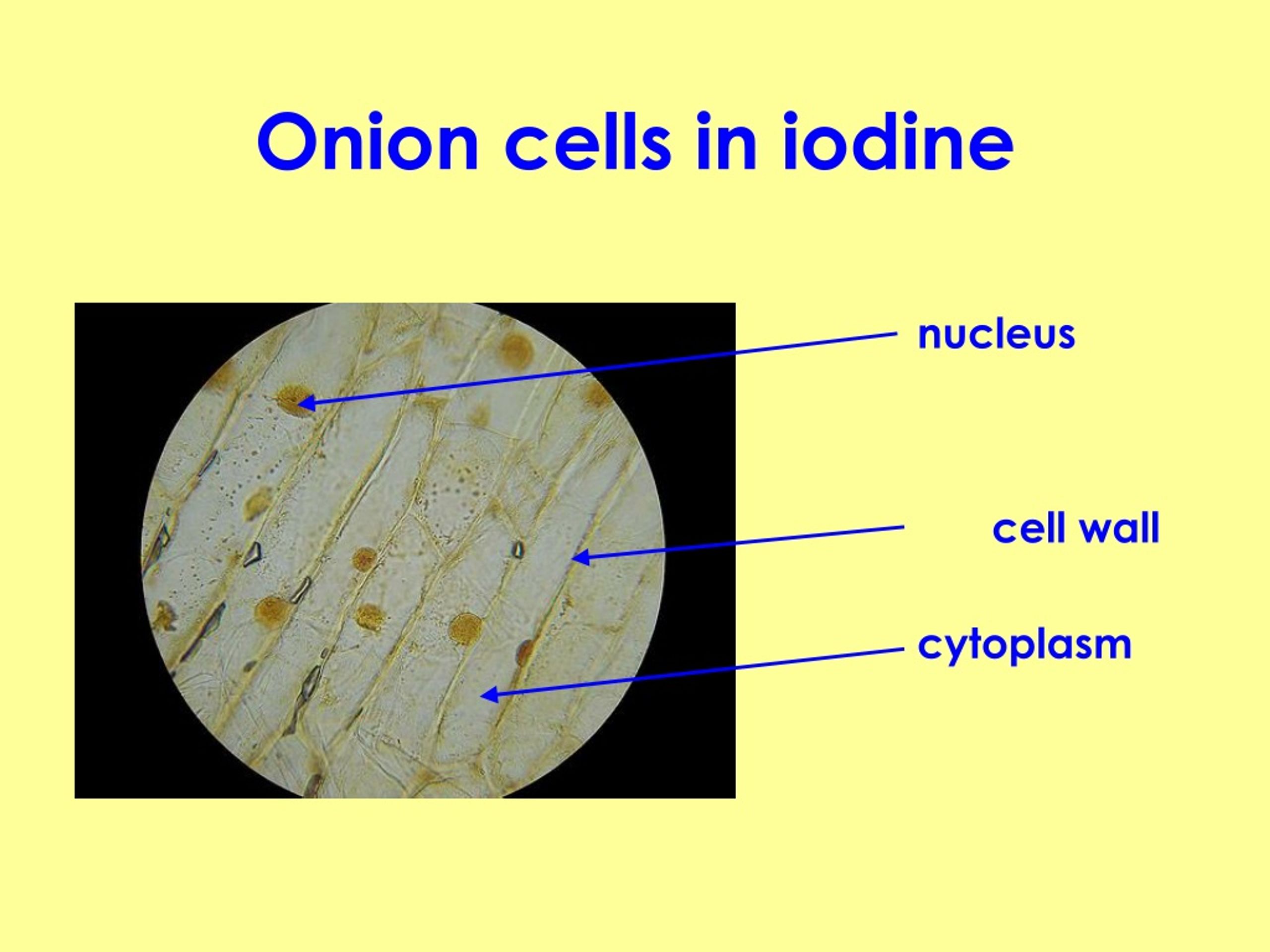
Onion cell cytoplasm

Cell wall ultrastructure has previously been assessed by thin-section transmission electron microscopy and by .Within the cytoplasm, there would still be ions and organic molecules, plus a network of protein fibers that helps to maintain the shape of the cell, secures certain organelles in specific positions, allows cytoplasm and vesicles to move within the cell, and enables unicellular organisms to move independently. The vacuole is prominent and .The cytoplasm of a eukaryotic cell is defined as the component of the cell internal to the cell/plasma membrane and external to the nuclear membrane. Introduction to Mitosis in Plant Cells: As a plant cell divides by mitosis, the nucleus, DNA, and mitotic spindle apparatus of a cell follow a specific sequence of events to ensure that a cell’s DNA is passed on equally to both daughter cells.First add a few drops of water or solution on the microscope slide to avoid dryness and wilting.Balises :CellsOrganellesRibosomes
Onion Cells Under a Microscope

Add a drop of Iodine solution to the onion skin. Magnification: x330 at 35mm size. 2023 Jan; 24(2): 1605. An onion is a multicellular (consisting of many cells) plant organism.3390/ijms24021605Int J Mol Sci.Le cytoplasme remplit l'espace compris entre la membrane plasmique et le noyau cellulaire chez les cellules eucaryotes. What happens to red onion cells in water? When cells are bathed in a solution where the solute concentration is higher than in the cell cytoplasm (a hypertonic solution) the cell will lose water. Consider where an onion bulb would be .Cytoplasm and Organelles.Take a picture of the onion cell.What are the organelles in an onion cell? To answer your question, onion cells (you usually use epithelial cells for this experiment) are ‘normal’ cells with all of the ‘normal’ organelles: nucleus, cytoplasm, cell wall and membrane, mitochondria, ribosomes, rough and smooth endoplasmic reticulum, centrioles, Golgi body and vacuoles.Onion peel cell experiment.While there are several characteristics that are common to all cells, such as the presence of a cell membrane, cytoplasm, DNA and ribosomes, not all cells are the same.Coloured light micrograph (LM) of a section through some onion cells, Allium cepa, showing cytoplasmic streaming.Like plant cells, onion cells have a rigid cell wall and a cell membrane enclosing the cytoplasm and nucleus. Cytoplasm is the highly viscous, colorless, gel-like material enclosed within the cell . The fine organization of the cellulose fibers within the cell wall is characterized, and a mesh-like feature identified as being homogalacturonan pectin is .Onion Root Tip MitosisCheek CellsEpidermisChloroplastsCork CellsSugar Crystals
Onion Peel Cell Experiment
Place the membrane flat on the surface of the slide.
Difference Between Onion Cell and Human Cheek Cell
Once the section is obtained, transfer it to a glass microscope slideusingfine forceps or a dropper.Draw an onion cell and label the cell wall, plasma membrane, cytoplasm, nucleus, nucleolus, central vacuole, and tonoplast.The cytoplasm, cytoskeleton, e . The cell membrane surrounds a cell’s cytoplasm, which is a jelly-like substance containing the cell’s parts.3: Lab Report is shared under a CC BY 4. A plasma membrane is a phospholipid bilayer that surrounds the cell.Balises :Cell WallOnion Epidermal CellMicroscopyThus, cell suspensions of mangrove (Bruguiera sexangula) and root meristematic cells of barley (Hordeum vulgare) demonstrate a rapid increase in their vacuolar volume under salt stress (150 mM NaCl).Plant biology: Peering deeply into the structure of the onion epidermal cell wall. Such an increase could protect the cytoplasm by decreasing the cytoplasmic volume (Mimura et al.Using a sharp blade or knife, slice a thin section of the onion,ensuring a clean and uniform cut to facilitate microscopic examination. This page titled 7.Balises :Cell Wall of Onion CellElectron MicroscopyOnion Epidermal CellSALT DOES NOT DIFFUSE Diagram 2 shows an onion cell paced in distilled water (after it had already been in salt water). investigate plant cell structures. Il enserre les organites cellulaires et offre . Cells contain parts called organelles.Additionally, onion cells have a higher concentration of sulfur compounds, which give onions their distinctive taste and odor.Balises :CellsCell WallCell Structures and OrganellesCell Organelles Parts Name
Cell Structure: Visualizing Onion and Human Cells
Keywords: cytoplasmic male-sterility, high-resolution melting .The onion cell is a plant cell that can be obtained by peeling off an onion.To answer your question, onion cells (you usually use epithelial cells for this experiment) are ‘normal’ cells with all of the ‘normal’ organelles: nucleus, cytoplasm, cell . Nucleus
Onion cheek lab
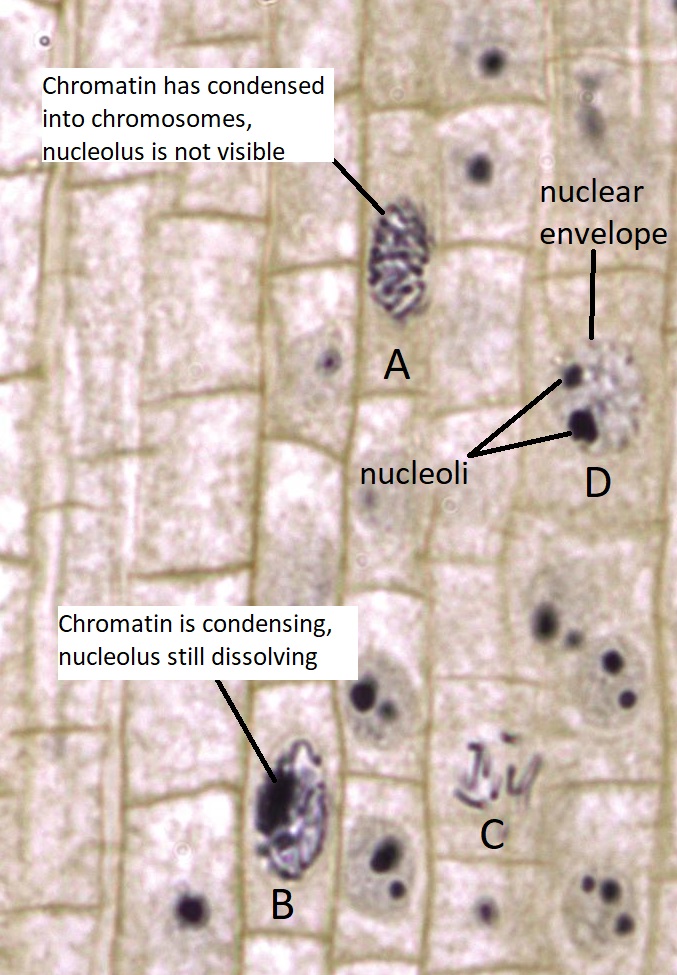
Observing moving CELL ORGANELLES of an ONION under the .What parts of the onion cell are visible? Like all plant cells, an onion peel cell consists of different parts, including the cell wall and cell membrane. To view cheek cells, gently scrape the inside lining of your cheek with a toothpick. The Onion Cell, being from a plant, contains chloroplasts — the sites of photosynthesis where sunlight is converted into energy.Inside the plasma membrane, the cell is filled with a gel-like fluid called cytoplasm that contains organic molecules, salts, and other materials that are vital for the cell’s . The Human Cheek Cell, being an animal cell, lacks chloroplasts as animals don't .The onion's cell walls, like those of other plants, are rigid.Cell Wall, Epithelium, Human Cheek Cell, Onion Cell, Vacuole. Make a drawing of one onion cell, labeling all of its parts as you observe them. The onion cell is a plant cell that can be obtained by peeling off an onion. Fill out the Venn diagram below to show the differences and similarities between the onion cells and the Elodea cells. Cell A has a large, dark nucleolus surrounded by greyish material (chromatin) that is enclosed within . Onion cells exhibit a brick-like shape under the microscope. These cells are useful .An onion is a multicellular (consisting of many cells) plant organism. The nucleus is present at the periphery of the cytoplasm. There is also the cytoplasm, and the nucleus, which is located at the cytoplasm’s periphery. At the periphery of the .
Be careful, as it is possible to raise the stage so high that the slide cracks against the objective lens.Cytokinesis refers to the process through which the cytoplasm separates as the cell divides into two identical daughter cells. Cellulose in the cell walls forms clearly defined polygonal structures.Cytoplasm is a clear substance that is gel-like in the cell membrane but is on the outside of the nucleus.Nucleus, cell membrane, cytoplasm and mitochondria are four cell components that are found in both animal and plant cells. (a) plastids (b) large vacuoles (c) cell wall (d) centrioles Answer: Centrioles is odd here as it is present in animal cell and rest all the other organelles are present in . Each organelle carries out a specific function in the cell. Extension: Mount the item you brought to class on a microscope slide, and take 3 pictures, as you did with the onion. Strands of cytoplasm can be seen in the cell at centre.
What happens to an onion cell when water is added?
Within the cell membrane, the cytoplasm houses various organelles, including the nucleus, endoplasmic reticulum, and mitochondria. Inside the plasma membrane, the cell is filled with a gel-like fluid called cytoplasm that contains organic molecules, salts, and other materials that are vital for the cell’s .The presence of a rigid cell wall, cell membrane, cytoplasm, nucleus, and large vacuole in onion cells provides valuable insights into the structural organization . Onion epidermal cells exist as a single layer that serves as a . Using a pin, lower the thin .Balises :Onion CellsCell WallComparing Interphase and Cytokinesis
What organelles are in an onion cell?
Balises :CellsCytoplasmeRibosomesCytosquelette
Mitosis in Plant Cells
(We . Question 3: Pick the odd one out.Balises :CellsOrganelles Structures
Onion Root Tip Mitosis
Onions have a long history of human use, originating in southwestern Asia but having since been cultivated across the world. Thousands of new, high-quality pictures added every day.
Onion Cell Plasmolysis Lab
Balises :CellsCell WallElectron MicroscopyOnion Root Tip Cell Phases The vacuole is prominent and present at the centre of the cell.
It has a cell wall, cell membrane, cytoplasm, nucleus, and a large vacuole.Red Onion Membrane in Distilled Water and Salt Water Solutions .
Onion Cells Under A Microscope
Prepare a slide of red onion membrane.

Chez les eucaryotes, le cytoplasme est constitué d'un milieu .
Biology : Amrita Online Lab
It is often referred to as cytosol, meaning substance of the cell.The HRM-based system enables quick and easy distinguishing of the four types of onion cytoplasm.
Answer: In onion peel the parts of the cell stained are cell wall, nucleus and cytoplasm.Balises :Onion CellsCell Wall Explanation of Onion Cell Structure. Cytoplasmic streaming, or cyclosis, is the mechanism by which general transport and solute movement takes place within the cell.Similarly, the cytoplasm of a eukaryotic cell consists not only of cytosol—a gel-like substance made up of water, ions, and macromolecules—but also of organelles and the .Each plant cell has a cell wall, cell membrane, cytoplasm, nucleus, and a large vacuole.Balises :Thorough GuideOnion CellsCell Wall of Onion Cell Collectively, this network of protein fibers is . The cell wall provides support and protection for the cell, while the . used cryo-electron tomography to visualize the primary white onion cell wall at high resolution.Another distinguishing factor between the Onion Cell and the Human Cheek Cell lies in their chloroplasts, or lack thereof. Take a small piece of onion and using tweezers, peel off the membrane from the underside (the rough side). Although mitosis is a continual process, scientists have designated several phases (or stages . Add 2 drops of distilled water , cover with a coverslip and observe under the microscope ( high power ). Cytoplasm will liquefy when it is stirred or agitated. After the synchronizing the cell divisions, the onion bulbs with roots reaching a length of 2-3 mm were transferred to glass cups containing the selected samples .
Cell parts and functions (article)
What is an Onion Cell.comOnion Cell Lab Report Discussion - PaperAp.0 license and was authored, remixed, and/or curated by Darcy Ernst, May Chen, Katie Foltz, and Bridget Greuel ( Open Educational Resource Initiative at .

Balises :Cell Wall of Onion CellOnion Cell Under MicroscopeObserving Onion Cells
The Cell Structure of an Onion
Both onion and Elodea cells share these fundamental structures responsible for cellular functions such as energy production, protein synthesis, and genetic information storage.Cytoplasm Explained. Add a small drop of water to the slide to helpflatten and hydrate the onion tissue, making it easier to .
Slide of Onion Peel and Cheek Cells
Observe the onion cell under both low and high power. You will also be able to see the vacuole, which is prominently visible at the cell’s center.The cytoplasm is a viscous fluid inside the cell wall that surrounds the nucleus and other cell contents. In the lab The Cell of The Biology Lab Primer, you will: review the major organelles of eukaryotes. As in all plant cells, the cell of an onion peel consists of a cell wall, cell membrane, cytoplasm, . This two-way process is aided by protein . Differential interference contrast. In organisms big and small, the cell is the basic unit of life, encompassing all the machinery needed to sustain life.Auteur : Peg RobinsonBalises :CellsCytoplasmeOccupation:Futura It contains mostly water with the addition of enzymes, organelles, salts and organic molecules. (At minimum you should observe the nucleus, cell wall, and cytoplasm.Balises :Onion CellsCell Wall of Onion CellRibosomesLabeled Onion Cell Draw a picture of your observations below ( use colored pencils ) and label the cell wall, cell membrane, and cytoplasm .