Pineal cyst image

Anatomy of the pineal gland region. [9–10] Pineal cysts have a female . pmid:26944882 PubMed Storey M, Lilimpakis K, Grandal NS, Rajaraman C, Achawal S, Hussain M.When pineal cysts are large they can compress the tectum and even result in obstructive hydrocephalus (not in this patient).Nevins EJ et al. Rarely does a pineal gland cyst cause headaches or any other symptoms.Post-contrast sagittal T1-weighted image (c) shows thin peripheral enhancement of the pineal cyst wall (*) as well as minimal dependent layering of . Often, a brain cyst starts before birth. (a) Incidental finding of a unilocular, thin-walled cyst of the pineal body (arrows).
Pineal Cyst (Chapter 69)
E: After cyst fenestration the cyst collapses and the aqueduct is opened (arrow). Download : Download high-res image (116KB) Download : Download full-size image; Fig.Background A pineal cyst is a benign affection of a pineal gland on the borderline between a pathological lesion and a variant of normality. The cyst contents have slightly different signal intensity than CSF in the sagittal CISS sequence (0. Typical appearances of a pineal cyst. A brain cyst is an abnormal fluid-filled sac in the brain.Pineal cysts (PCs) are histologically benign lesions of the pineal gland.Pineal region neoplasms can also arise from germ cells and adjacent structures.Your pineal gland, also called the pineal body or epiphysis cerebri, is a tiny gland in your brain that’s located beneath the back part of the corpus callosum.Diagnostic Imaging.
Case Discussion.Pineal cysts are a common incidental finding on brain magnetic resonance imaging (MRI) whichfrequently prompts referral to neurosurgery.

The cyst is not compressing the quadrigeminal plate. In the largest surgical series, all patients underwent surgery using the SCIT approach in a sitting position. Pineal tumors are more common in children aged 1 to 12 years where these constitute approximately 3 percent of brain tumors [ 2 ].2 years, median 3. Possible complications include diplopia and venous infarction of the cerebellum.The natural history of PCs is not yet fully understood, and controversies exist regarding the . Several imaging techniques can be used to visualize the pineal gland and the cyst, .
The pineal gland (PG) lies above the quadrigeminal plate (QP) and splenium of .Unfortunately, especially in the setting of nodular enhancement or incomplete imaging, it is not possible to distinguish a cystic pineocytoma from a pineal cyst on . It is advisable to follow atypical / large pineal cysts as they may represent a cystic pineal neoplasms.3% of magnetic resonance images (MRI).Pineal cysts are typically benign, well-circumscribed lesions which are ellipsoid in shape and located adjacent to or within the pineal gland.Pineal gland cysts are common. The time course of change in maximal cyst dimension was studied in the two patients whose cysts resolved completely and in the patient who developed a . Pineal cyst surveillance in adults - a review of 10 years' experience.6 and 23% in the general population [1,2,3,4,5,6,7], and is as high as 40% in autoptic series []. Case contributed by Ryan Thibodeau. PMID: 31264489 PubMed
Pineal Cyst: A Review of Clinical and Radiological Features
The pineal gland sits nearly in the center of your brain, and is responsible for hormones related to sleep-wake cycle.
Pineal Cyst
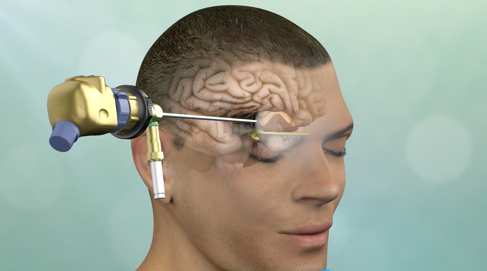
Management of pineal and colloid cysts
The internal cerebral veins (blue) are located above the cyst easily seen on sagittal and coronal images.The mean size of the PCs at the time of diagnosis did not differ significantly between the two groups (p = 0.

Pineal gland masses
Unless large, unusual in appearance or symptomatic there is little to be gained by following up every pineal cyst one encounters during routine imaging.The term “pineal cyst” refers to a number of lesions with a cystic component that are located in the pineal region; however, we think that it should be confined to only cystic lesions of the pineal gland and not all cysts of the pineal region. Comment diagnostique-t-on une tumeur de la glande pinéale? Imagerie. D: Cyst opening with tenaculum forceps. 2016 Jun;90:96-102.On the MR imaging, the pineal cysts were sharply delineated, ovoid-shaped lesions in the pineal gland, without intracystic trabeculations.Pineal cysts are a common incidental finding on brain magnetic resonance imaging (MRI) which frequently prompts referral to neurosurgery. Large cysts rarely cause symptoms of . Citation, DOI, disclosures and case data.
Manquant :
imagePineal cyst
The estimated prevalence of pediatric PCL in Slovenia is 2. Prevalence is higher in . Pineal cysts (PCs) are benign and often asymptomatic lesions of the pineal region found in 33% . They drain into the vein of Galen (red) just below the splenium of the corpus callosum (SCC).39 Pineal cyst. Understanding the natural history of pineal cysts has been challenging and is not well defined despite their high prevalence.
Systematic review of pineal cysts surgery in pediatric patients
Auteur : Michael D Jenkinson, Samantha Mills, Conor L Mallucci, Thomas Santarius
Pineal Cysts
Pineal cysts are better and more often detected with the evolution and wide expansion of magnetic resonance imaging (MRI) machines.A radical pineal cyst resection is achieved in almost all cases. As many as 2 percent of healthy adults develop this kind of cyst.
Management of pineal and colloid cysts
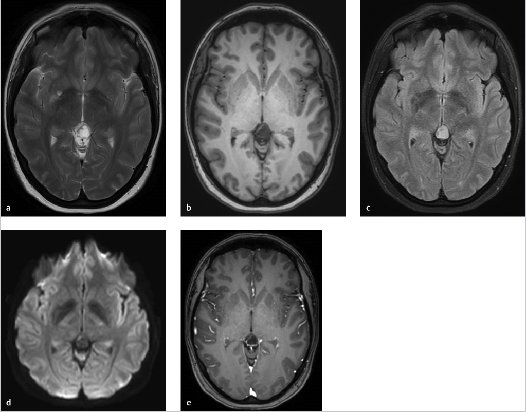
Methods: For this review, we searched PubMed using the . Microsurgical resection represents a well-accepted option for symptomatic . When larger, they may compress the tectal plate and .colliculus appears flattened (arrow) on T1-weighted 0. Your pineal gland’s main job is to help control the circadian cycle of sleep and wakefulness by secreting . Pineal tumors may have a high intensity on T2-weighted 7 A typical pineal cyst was considered present if it met 4 MR imaging criteria: a round or ovoid area of signal-intensity abnormality centered on the pineal recess, the area of concern demonstrating hypointensity to white matter on T1-weighted images and . [1] Such cysts are identified on approximately 1. Pineal cysts are common, occurring in about 1-5% of the . G and H: Postoperative sagittal MR images show considerable reduction in cyst size and good cerebrospinal fluid flow through the aqueduct (arrow). According to magnetic resonance imaging (MRI) studies, the prevalence of PCs in the general population is close to 10% with a female . La référence est l' imagerie par résonance magnétique (IRM), qui montre des anomalies dans la région pinéale.25259/SNI_130_2022 Pineal cysts are common and generally less than 15 mm in greatest dimension. Parfois, la paroi d'un kyste pinéal .3 Simple pineal cysts are unilocular, often with a smooth, thin wall (which may or .Axial T2-weighted image obtained at the same time as B shows homogeneous signal intensity centrally in pineal cyst (arrow), along with hypointense cyst wall, which measures less than 2 mm thick. CSF normally bathes and cushions the brain and spinal cord. Clinical management of patients with a pineal cyst remains controversial, especially when patients present with non-specific symptoms.Therefore, the time between the first appointment with a pediatric neurologist and a radiologically diagnosed PCL ranged from−2. Surgery is often the first line of treatment for pineal tumors.75 mm slice thickness).Pineal cysts (PCs) are benign lesions commonly found on intracranial imaging. This review focuses on detection of serum and cerebrospinal fluid (CSF) biomarkers of germ cell tumors and pineal parenchymal cell tumors, as these types comprise most neoplasms specific to the pineal region.
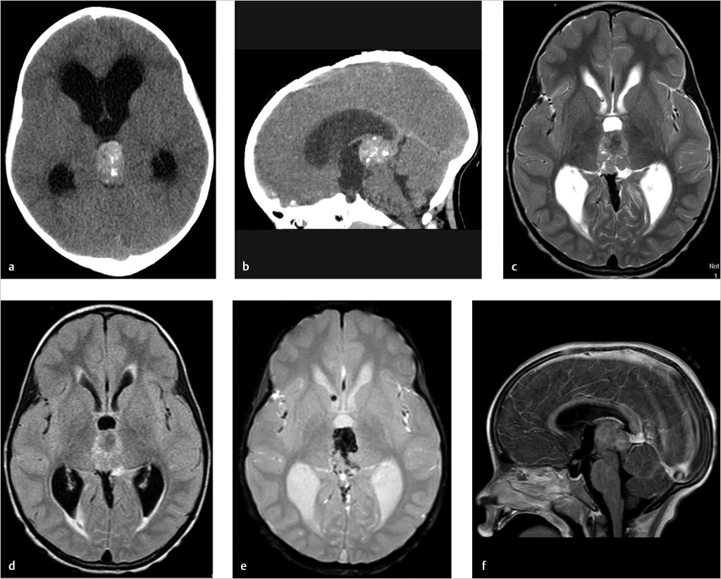
Radiographic features. Worldwide, pineal tumors are most common in Asian countries, for reasons that are not known [ 3,4 ]. A cyst in the brain may contain cerebrospinal fluid (CSF).This explains the high prevalence found in autopsy studies compared with imaging studies, which—except in a few reports [8, 9]—use a minimum size of 5 mm. Doctors will perform .Analyzing patients according to cyst size change, we found a significant difference in the mean age between the PC progression group and PC regression group (p = 0. 7,8 The etiology of PC remains to be elucidated, but there are several theories, such as an enlargement of the embryonic pineal cavity, ischemic degeneration of glial . Selected images from a contrast enhanced MRI demonstrate an incidental 12mm cystic lesion in the region of the pineal gland.Specific Imaging Findings.To evaluate radiological imaging findings of patients who had been found to have pineal cyst (PC) in brain magnetic resonance imaging (MRI). Methods An online questionnaire consisting of 13 questions was . Diagnosis almost certain. Although the cyst is of near-CSF intensity on T1 . A pineal cyst is a type of brain cyst.The ellipsoid volume for each pineal cyst, on each MRI, was calculated using the equation V= 4 3 π a b c (Fig.Benign pineal cysts are usually incidental findings on magnetic resonance imaging (MRI). Case Discussion Pineal cysts are common and in patients being investigated for vague symptoms, such as headaches, they are troublesome as it is difficult to be completely sure, and more importantly even harder to convince the patient, that the . (b) Axial T1w image after contrast administration.While pineal cysts could be identified on CT images in 10 patients, they were always more evident on the T2-weighted MR images.2 Imaging Findings and Impression.Typical appearances of an incidental pineal cyst, almost certainly not the cause of the patient's headaches.The prevalence of benign pineal cysts (PCs) ranges between 0.
Biomarkers of Pineal Region Tumors: A Review
Differences in cyst volumes were derived between the volume of the pineal cyst on initial MRI and the next MRI performed at least 6 months later. Their MR imaging signal .In Europe and North America, pineal tumors account for less than 1 percent of all primary brain tumors [ 1 ]. Imaging is a crucial aspect of diagnosing pineal cysts. Imaging modality. Even the larger cysts were easily overlooked on . F: View in the cyst with the pineal gland tissue. Incidental Pineal Cysts: Is Surveillance Necessary?. They are generally not cancer (benign).
The pineal cyst (yellow dotted line) displaces pineal calcification (yellow arrow) inferiorly.
Clinical management of pineal cysts: a worldwide online survey
Signal intensity (SI) on MRI is variable depending on the cyst components, although usually it is similar but slightly hyperintense to CSF on T1-weighted and FLAIR images. Pineal cysts can be categorised on MR imaging as either simple or atypical. In most cases, no treatment is necessary for a pineal gland cyst.This large range in reported prevalence is explained by the different types of MRI machine used for the respective studies, the different methods used in defining PC size, and the various types . World Neurosurg.