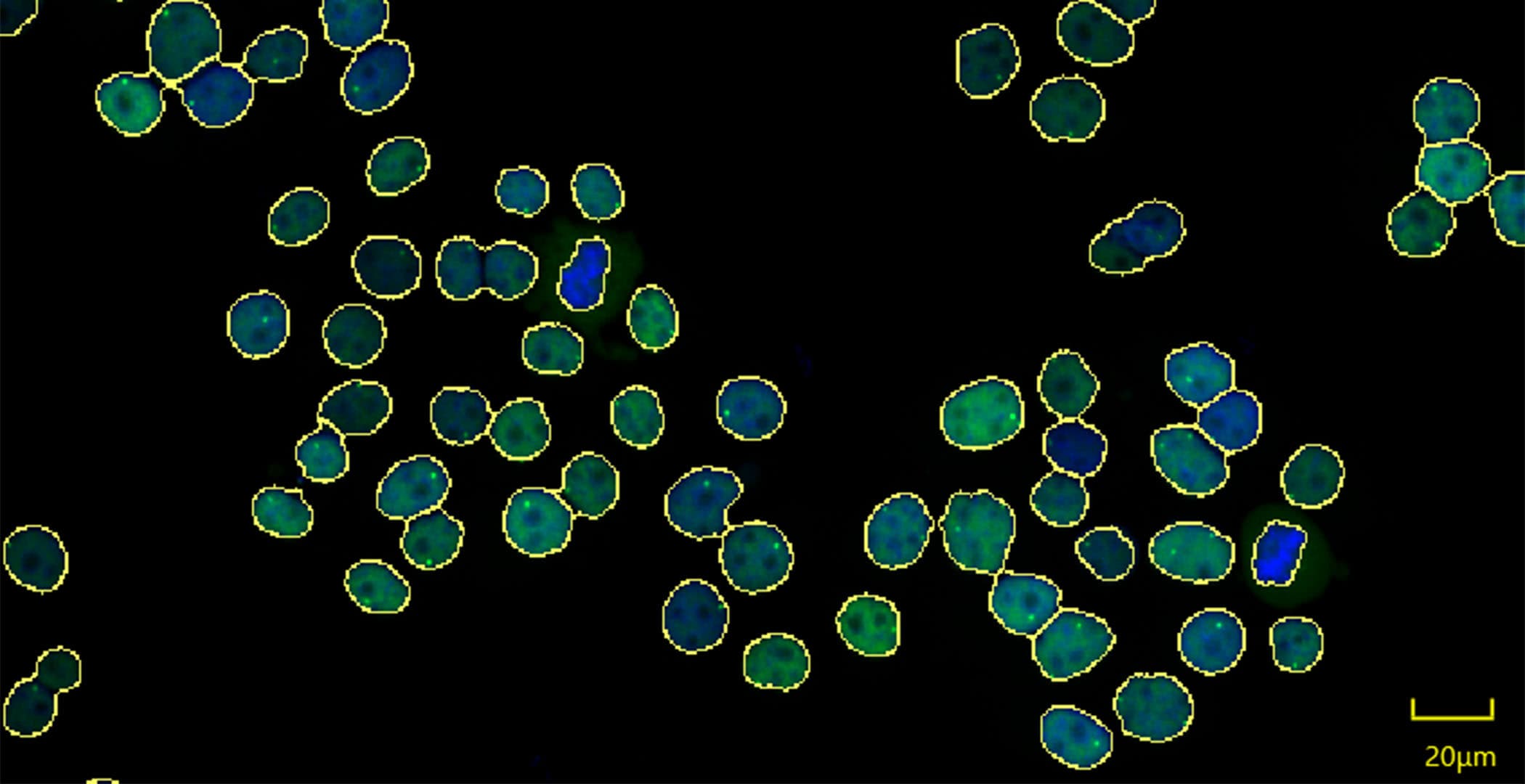
Single cell imaging

The Au nanoparticles (AuNPs) were .Single-cell technologies have revealed the complexity of the tumour immune microenvironment with unparalleled resolution 1,2,3,4,5,6,7,8,9.0-fold higher signal at room temperature compared with ilux .Especially for single cell projects, the long-term monitoring of cell growth, differentiation, and drug effect analysis is of great significance., the most abundant cellular proteins .
Recent advances in mass spectrometry imaging of single cells
Single cell imaging methods like fluorescence microscopy have made it possible to acquire cellular features like morphology or cell area at such a high .
Single-Cell Imaging of Metastatic Potential of Cancer Cells
Single-cell image analysis provides a powerful approach for studying cell-to-cell heterogeneity, which is an important attribute of isogenic cell populations, from . Yet, a major hurdle has been the lack of facile and predicative preclinical animal models that permit dynamic visualization of T cell immune responses at single-cell resolution in vivo.Le LabTech Single-Cell@Imagine regroupe les compétences humaines, technologiques et les équipements permettant d’analyser l’expression de milliers de .
Statistical integration of these datasets identifies a role for KLK12 in motility at NEUROG3 expression onset, while also providing a methodology . The new facility has design specifications aimed at spatial resolutions below 50 nm, with a variety of techniques .Here, we developed a method to study cell:cell firm adhesion under shear-stress conditions coupled to high-content live-cell imaging, and single-cell RNAseq analysis.
CosMx SMI Overview
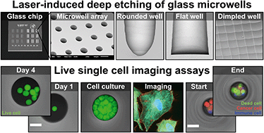
Applications based on deep learning techniques in the field of single-cell optical image studies are reviewed, which include image segmentation, super-resolution image .The experimental and computational tools that enable continuous imaging of single cells for days and weeks have advanced rapidly in recent years, and solutions to . Single-cell multimodal . It is the flexible, spatial single-cell imaging platform that will . Materials and .A 3D illustration of pancreatic cancer cells. Nature 480 , 139–141 ( 2011) Cite this article.Single cell live imaging allows for time-series understanding of individual cells while maintaining their culture or living conditions. ( 2021) 17: e9653. Cell And Tissue Culture49.Single-Cell Imaging Technique. The major building block molecules of a cell comprise proteins, nucleic acids, lipids as well as phospholipids, and carbohydrates among others.On the basis of a comprehensive proteomic map of the mammalian cell line, U2OS, reported by Beck et al.Single‐cell imaging of genome organization and dynamics.Single-cell technologies have enabled the characterization of the tumour microenvironment at unprecedented depth and have revealed vast cellular diversity among tumour cells and their niche. Methods: We used cutting-edge multiplexed ion beam imaging by time of flight to compare pulmonary arteries (PAs) and adjacent tissue in PAH lungs (idiopathic [I]PAH and HPAH) .

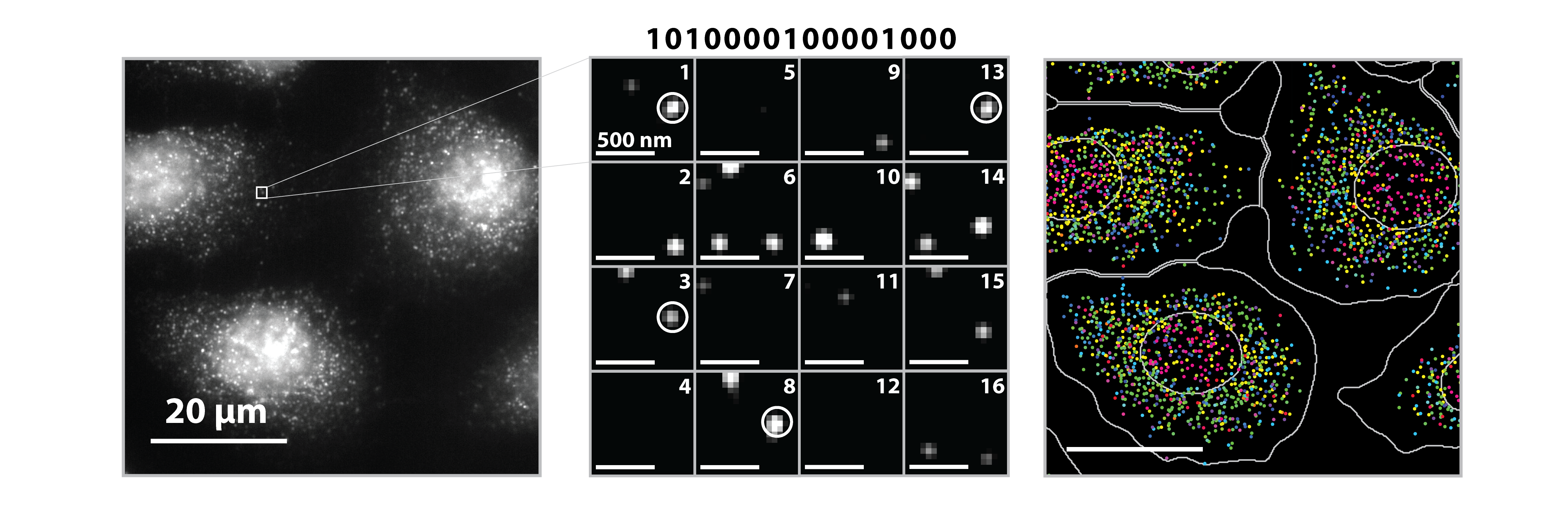
Both spontaneous and stimulated emission routes are a transition form of released energy for excited state [].What is Single-Cell Imaging? | NanoString. characterize the differentiation dynamics of single human pancreatic progenitors into endocrine cells in 2D and 3D models by RNA sequencing and live imaging.Beydag-Tasöz et al. As a nonscanning imaging technique, light field microscopy (LFM) is a critical tool .In imaging, single-molecule fluorescence resonance energy transfer . Using genetically directed bioluminescence from AVP or VIP neurons in the SCN circadian pacemaker, they unravel distinct functions of the circadian clock in . Single-cell imagers use sequential cycles of probe hybridization and imaging and offer the potential to combine the benefits of scRNA-seq analysis with added spatial resolution at single-cell or even subcellular resolution.Using image-based single-cell data, the authors map genetic interactions and cell morphologies of more than 6,800 genes in Drosophila cells and use machine learning to predict gene functions and modules.Raman spectroscopy can provide nonbiased single-cell analysis based on the endogenous ensemble of biomolecules, with alterations in cellular content indicative .

Multimodal volumetric imaging at the single-cell resolution in intact human tumours can provide unique molecular and pathophysiological insights into how solid tumours are formed, which may be .Objectives: This study aims to elucidate immune-driven vascular pathology by identifying immune cell subtypes linked to severity of pulmonary arterial lesions in PAH.Little did he know . A team in the U.Surface plasmon resonance microscopy (SPRM) is a versatile platform for chemical and biological sensing and imaging.Most clinical strategies rely on histopathological . Expression of ilux from pQE (−) in Top10 cells resulted in a 2.Visualizing molecular distribution at the single-cell or even sub-cellular level can provide us with richer cell information, which strongly contributes to advancing . Next we asked if the IMP-Y1764 probe retains its ability to detect the metastatic potential of tumor cells despite evolving signaling programs and tumor . Confocal microscopy and STED microscopy are based on point‐scanning and thus are slower.The single cell imaging facility is designed around OM52 compact lenses capable of operating in a variety of high demagnification configurations including the spaced Oxford triplet and the double crossover Russian quadruplet.Single-cell analysis is not confined to’omic analytes, either.UC Santa Cruz researchers have developed a method to solve this by building a microscopy image generation AI model to create realistic images of single .Single-cell analysis: Imaging is everything.Single-cell imaging using radioluminescence microscopy demonstrated unexpected preferential accumulation of [18 F]HFB in fragments of membranes from dead cells, highlighting the importance of radioluminescence imaging for accurate characterisation of cell labelling efficiency in vitro prior to in vivo models.We report here the development of coreactant-based electrogenerated chemiluminescence (ECL) as a surface-confined microscopy to image single cells and their membrane proteins.CosMx SMI enables rapid quantification and visualization of up to 6,000 RNA and 64 validated protein analytes. Nascent intron spots are .

Great progress in exploring its applications, ranging from single-molecule sensing to single-cell imaging, has been made.

Wide field microscopy including those used in SMT is suited to probe the fast dynamic events reaching milliseconds resolution. Herein, we developed a dual-modal microscopy imaging strategy to For spatial multi .Here, the authors apply live-cell and in situ fluorescence imaging at the single-molecule level to examine lambda DNA replication in single cells, finding that .In order to elucidate the demand single-cell analysis places on mass spectrometry detection, we have estimated the abundance of proteins and lipids in a single mammalian cell (Table 1).
Tools and methods for high-throughput single-cell imaging
Integrating single cell omics and single cell imaging allows for a more effective characterisation of the underlying mechanisms that drive a phenotype at the tissue level, .Here, we discover the stimulated emission route of ECL and thus establish a reversible tuning ECL microscopy with a red-shifted beam for single-cell imaging (Fig. T cell immunotherapies have revolutionized treatment for a subset of cancers. Intron sequential fluorescence in situ hybridization (seqFISH) (top left) can visualize the nascent transcriptome (>10,000 genes) with single RNA molecules in the nucleus of a mouse embryonic stem cell (mESC) ( 3 ).80 results for Single-cell Imaging Concepts identified: Technique: Single-cell Imaging. Breast cancer is the most . Liangqi Xie and Zhe Liu Author Information. Ryan Thiermann, Michael Sandler, Gursharan Ahir, John T. Single-cell nuclear architecture . These data reveal how a Cdk2 and Cop9 signalosome interaction affects hemocytic cell immunogenicity by triggering senescence .Single-cell SERS imaging of dual membrane receptors on HeLa cells cultured on substrates with different stiffness. However, the way in which cisplatin-induced DNA lesions regulate interactions between transcription factors/cofactors and genomic DNA remains unclear.Single-Cell Imaging of Signaling through the GIV-PI3K Axis Maintains Its Usefulness in Tracking Metastatic Potential Despite Tumor Evolution and Onset of Chemoresistance.Single-cell volumetric imaging is essential for researching individual characteristics of cells.Single-cell sequencing datasets are key in biology and medicine for unraveling insights into heterogeneous cell populations with unprecedented resolution.The difference is that spontaneous emission happens without .
Single-cell multimodal imaging uncovers energy conversion
Single-cell imaging and analysis are fundamental to research biomedical mechanisms, which create a comprehensive understanding at the cellular level. August 2, 2022.Cisplatin is an extremely successful anticancer drug, and is commonly thought to target DNA.IN VIVO SINGLE CELL IMAGING. generate a Cre-inducible mouse reporter with green and red luciferases fused to PER2, enabling cell-specific circadian measurements with a dual-color imaging device.Whereas cell cycle typically takes ~1 day, differentiation typically lasts a few days or even weeks. Publication Year.Tools and methods for high-throughput single-cell imaging with the mother machine.Purpose The absence of clinically applicable imaging techniques for continuous monitoring of transplanted cells poses a significant obstacle to the clinical .
Inauguration du Single-Cell@Imagine
In order to achieve effective and continuous cell imaging, super resolution fluorescent microscopy has become a widely used imaging tool [8-10].
Imaging nuclear architecture in single cells
Auteur : Mojca Mattiazzi Usaj, Clarence Hue Lok Yeung, Helena Friesen, Charles Boone, Brenda J Andrews
Imaging nuclear architecture in single cells
The major technological themes of this review include (1) single-molecule imaging in vitro, in-cell lysate and live cells at high resolution, sensitivity, throughput, and biocompatibility; (2) in-cell single-molecule labeling of genomic DNA loci, mRNA transcripts and nascent .
Methods and applications for single-cell and spatial multi-omics
Scientists can see inside a single cancer cell
Although single-cell and ‘omic technologies are relatively young fields that have been developing the past 20 years [], single-cell imaging in the form of microscopy can be traced back to the 1600s, when Robert Hooke first described the small rectangular compartments he observed with his microscope in cork bark as ‘cells' []. 1,2 Two-dimensional single-cell imaging based on conventional wide-field fluorescence microscopy has been extensively employed to visualize the shape and detailed inner structures. Here, optically clear immunocompromised zebrafish were engrafted . Exscientia, of Oxford, UK, is throwing its weight behind another foundational technology: high-throughput single-cell imaging. Home » Blog » What is Single-Cell Imaging? Categorized As: CosMx SMI Multiomics Spatial Analysis.Researchers have developed a method to use an image generation AI model to create realistic images of single cells, which are then used as 'synthetic data' to train .Single-cell multimodal imaging uncovers energy conversion pathways in biohybrids | Nature Chemistry.
In vivo molecular and single cell imaging
These advances allow . A recently developed technology for spatial single-cell imaging, the CosMx Spatial .Single-cell nuclear architecture captured by multiplexed genomic and transcriptomic imaging.To obtain a higher brightness for imaging of single E.
Single‐cell imaging of genome organization and dynamics
Sauls, Jeremy W. Published: 27 July 2023.


