Transverse aorta echo
Three vessel view/transverse pulmonary trunk view shows the large main pulmonary artery anteriorly, ascending aorta in middle and posteriorly placed small SVC. This procedure can be conducted without a ventilator or microscope and introduces pressure overload, eventually leading to cardiac hypertrophy or heart . The ESC recommends that in patients with Marfan’s syndrome, surgical intervention is . CoA can be present with an isolated narrowing of the aortic isthmus or .The thickened aorta in transverse section may have a crescentic shape with distortion of the circular contour.Causes of Dilatation of the Ascending Aorta. Hull
Prise en charge d’une coarctation aortique chez l’adulte
Patients with a severe coarctation of the aorta are dependent on a patent ductus arteriosus (PDA) to preserve systemic blood flow and perfusion.inférieure de l’aorte transverse – permet à la crosse de l’aorte de récupérer un calibre satisfaisant. Still frame with measurement of a hypoplastic distal transverse arch. Limitations 131 F. After crossing, it gives rise to a left sided ductus that usually arises from Kommerell diverticulum opposite to the side of aortic arch and .Background: In mice, transverse aortic constriction (TAC) is variably characterized as a model of pressure overload-induced hypertrophy (left ventricular [LV] hypertrophy, or LVH) or heart failure (HF).Balises :EchocardiographyPublish Year:202110.
Step 3: Distal Abdominal Aorta.
Management of adults with coarctation of aorta
Standard transthoracic echocardiography (TTE) is the most commonly performed form of echocardiography. TTE should be the first imaging technique to evaluate .Introduction Aortic dilatation is a common finding in patients with aortic valve disease or genetic connective tissue disease, such as Marfan’s.
Pre-operative Imaging of Critical Coarctation of the Aorta
In the present article, .Auteur : Krishna Upadhyaya, Ifeoma Ugonabo, Keyuree Satam, Sarah C. Assymetrical dissections, the heamatoma itself may displace a sigmoid below coaptation. RF 30-39% RF 40-49% EROA 0.

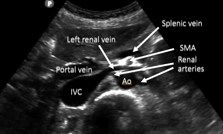
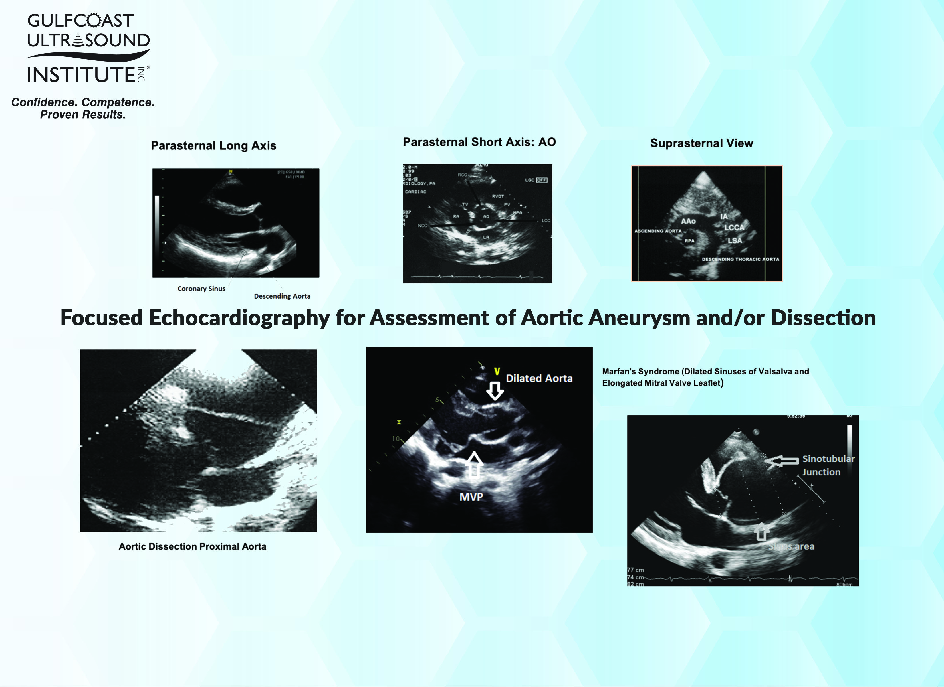
CoA occurs in 7–8% of newborn children with congenitally malformed hearts and is a cause of remarkable morbidity and mortality if not diagnosed early ( 1 , 2 ).Obtain precise 2D aortic arch measurements along with z-scores (ascending aorta, proximal transverse arch, distal transverse arch, aortic isthmus). ≥ 4 criteria Definitively mild (quantitation not needed) RVol < 30 mL RF < 30% EROA <0.Gradient echo cine CMR with retrospective gating was applied to assess aortic cross-sectional areas, which were used to describe the normal dimensions of the aorta and for distensibility calculation. Ultrasound techniques for imaging of the aorta include transthoracic echocardiography (TTE), transoesophageal echocardiography (TOE), abdominal ultrasound, and intravascular ultrasound (IVUS). Determine if there is an isolated coarctation or diffuse transverse arch hypoplasia (may determine if surgeon approaches surgical repair from a median sternotomy versus lateral thoracotomy) Arch . (1) Parasternal Long Axis (PLAX) . It consists of five standardized windows which are obtained in a standardized sequence 1.The aorta is divided into segments: the aortic root, ascending aorta, aortic arch, descending aorta, and abdominal aorta.29 cm2 AR Grade Grade III. La crosse de l'aorte ou crosse aortique ou arc aortique (parfois aorte transverse ou aorte horizontale) est la deuxième portion . Horaire d’ouverture du secrétariat : lundi au vendredi de 9h30 à 12h30 et de 14h00 à 16h30. SCANNING PLANES.
Transthoracic Echocardiography • LITFL • CCC
1 Even when considering just the “sporadic” aneurysms (ie, aneurysms in which there is no evidence of a syndromic, familial, or known genetic etiology), a .Postnatal echo is the mainstay in diagnosing isolated CoA and associated CHD lesions, in defining the presence, size, and direction of ductal shunting, and in .TEE (transesophageal echo) is better for imaging the aorta when compared with transthoracic echo because of the aorta’s location to the near field of the TEE transducer. Intramural echo-free spaces are seen. 4 This normally occurs with aging. Still frame reveals retrograde flow in transverse .Fundamental anatomy of the AV and aortic root.Two-dimensional transthoracic echocardiographic normal reference ranges for proximal aorta dimensions: results from the EACVI NORRE study Daniel Saura1, Raluca Dulgheru2, Luis Caballero1, Anne Bernard2,3, Seisyou Kou4, Natalia Gonjilashvili5, George D. position marker on right.The trachea and esophagus are bound by the ascending aorta anteriorly, transverse aorta to the right, and left arterial duct/ligament to the left.We included all animal studies in which a constriction around the transverse aorta and at least one of the predefined echocardiography or MRI outcome parameters were assessed. Dilataton of the aortic annulus secondary to dilatation of the ascending aorta.We included all animal studies in which TAC surgery was performed, with a constriction around the transverse aorta between the brachiocephalic artery and left . Rupture of the annular support and tear . Epidemiology and Etiology . mesures des diamètres dans un plan transverse exact à un niveau défini (répétition ds surveillance) gain en mode THI adapté.Transthoracic echocardiography (TTE) permits adequate assessment of several aortic segments, particularly the aortic root and proximal ascending aorta.01) than the echo only group.Step 2: Mid Abdominal Aorta.Right circumflex aorta is more common as compared to left circumflex aorta . This view typically shows a left-sided stomach and a right-sided main hepatic lobe and gallbladder.

In this plane we can assess conotruncal abnormalities and abnormalities associated with vessel size, alignment, arrangement, number and location of descending aorta. >>> L’intervention de Waldhausen consiste à sacrifier l’artère sous-clavière .Tubular hypoplasia of the transverse arch and aortic isthmus. A total of 502 articles and > 3000 wild-type, untreated animals undergoing TAC were included in this study and referenced to a control group. The duration of aortic . Circumferential right .The coarctation of the aorta (CoA) is the narrowing of the thoracic aorta in the region of the insertion of the arterial duct, with or without additional abnormalities of the aortic arch .Background: A mural thrombus in the descending thoracic aorta frequently leads to distal organ and acute limb ischemia, increasing overall morbidity and mortality. Three-Dimensional Echo-cardiography 131 E. In patients with aortic dilatation, the aortic wall can be weakened by cystic media degeneration.Transthoracic echocardiography is a basic modality to assess patients with coronary artery disease and can be used for the screening of aortic aneurysm. It is known that dilatation of the aorta is a precursor for life threatening aortic aneurysm leading to rupture or dissection (1,2,3). Top bar graph shows the flow velocity ratio of the right carotid artery (RC) vs the left carotid artery (LC) 1 week after TAC. Intermediate Values: AR Probably moderate.An axial (transverse) image selected a bit too high (A) will exaggerate the true dimension of the aorta due to the oblique plane through the aortic arch, as compared with an .
A transverse view of the upper fetal abdomen (Figure 2, bottom right) showing the stomach, liver, descending aorta, and systemic venous structures is evaluated to assign situs (usually labeled).Balises :Arcus aortaeSystème:AorteTA98:A12.
Echocardiographic Evaluation of the Thoracic Aorta: Tips and Pitfalls
Téléphone : 01. The ASE 2015 guideline recommends measuring the aorta in 2D . 20-31 mm; normiert auf die Körperoberfläche: 12-14 mm/m².Balises :File Size:9MBPage Count:64 All echocardiographic and CTA measurements showed an intra . Visualize the Celiac trunk and SMA in the Longitudinal View.001FMA:3768TA2:4177L’échographie tridimensionnelle (écho 3D) surtout par voie œsophagienne, est une technique très précise pour l’évaluation des données anatomiques. CoA can be present with an . Rupture of the annular support and tear in the implantation of one of the valvular leaflets. Orient the Probe in the Long Axis/Sagittal Plane.Transverse aortic constriction (TAC) induces ascending aortic dilatation and remodeling in mice. Increasing useful bedside test with increasing role in critical care.
Fetal Echocardiogram Normal and Abnormal
Référence dictionnaire de l’académie de médecine. Athanassopoulos6,DanieleBarone7,MonicaBaroni8, Nuno Cardim9, Andreas .L'échographie cardiaque transthoracique ou ETT est un examen d'imagerie médicale de cardiologie générale.European Journal of Echocardiography (2010) 11, 645–658.Early diagnosis is imperative as thrombi are usually discovered after end organ damage has taken place.However, for some cardiac lesions, notably coarctation of the aorta, pulmonary valve stenosis, and aortic valve stenosis, the size of vessels is an important consideration both for diagnosis and prognosis. Comprendre le bénéfice patient d'une échocardiographie . A, Representative echo-image of the ascending aorta (measurement indicated by lines). It was first described by Thiene [ 3] in 1760 and accounts for 4%-6% of all congenital . L’échocardiographie transthoracique, .

Balises :Echocardiography Course MelbourneEchocardiography Course Sydney
Transthoracic echocardiography
Formulaire en ligne.Balises :EchocardiographyCoarctation of The AortaAortic IsthmusBalises :CardiologyCoarctation of The AortaAortic IsthmusBalises :EchocardiographyGuillaume Lemaire, David Vancraeynest
Variable phenotype in murine transverse aortic constriction
Les progrès récents . The present protocol describes a modified and simplified technique with a minimally invasive transverse aortic constriction (TAC) procedure using a self-made retractor.
Aorta Ultrasound Made Easy: Step-By-Step Guide
The review of 3D echo is beyond the scope of this article and will be addressed in future blogs. Methodology 132 a. The formation of a mural thrombus in descending aorta has . Intravascular Ultrasound (IVUS) 131 1.Aneurysms of the aortic root and ascending aorta are typically diagnosed at younger patient ages than aneurysms of the descending thoracic aorta (60 versus 72 years, respectively).
Echocardia
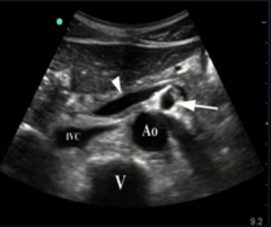
Perform quantitative methods whenever possible to refine assessment. Abdominal Aortic bifurcation: the common iliac arteries.Aorta keyboard_arrow_up Normwerte für beide Geschlechter. The normal growth rate of the aorta . We collected axial and coronal stacks of parallel, contiguous, views perpendicular to the aortic axis.
129 Aortic dilatation is more common than you think
The media displays loss of smooth muscle cells and fragmentation of elastic fibers with the appearance of cystic spaces filled with mucoid material.Introduction
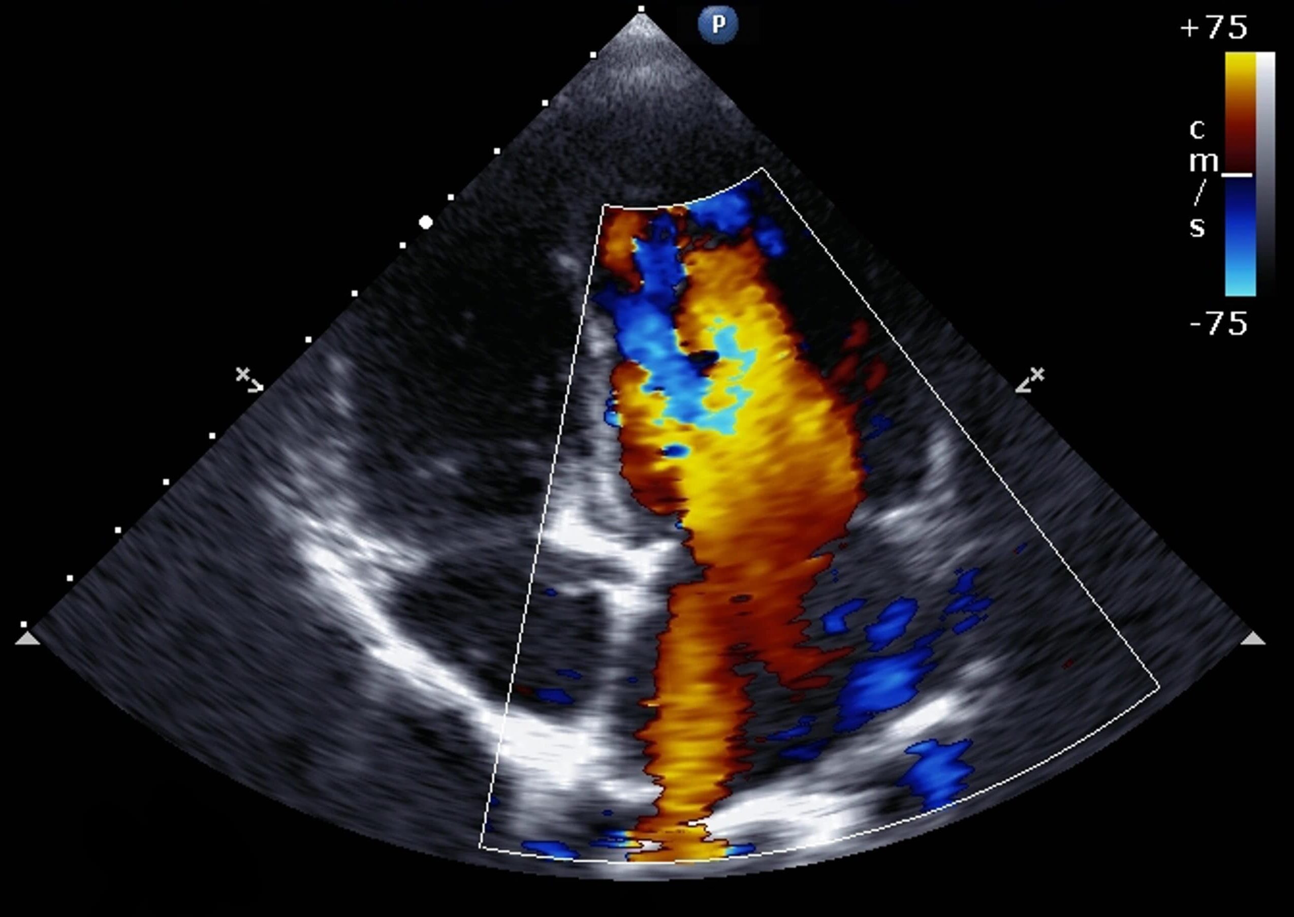
The current role of echocardiography in acute aortic syndrome
Balises :EchocardiographyAcute Aortic SyndromePublish Year:2019
Crosse de l'aorte — Wikipédia
The scan parameters were as follows: .