Two photon excitation microscopy
The comparison between single .
In 1931, the theoretical basis of two-photon excitation was established by Maria Goppert-Mayer, and this photophysical effect was . Checkforupdates. It also produces higher-contrast images and is a novel method to trigger localized photochemical reactions.Two-photon microscopy excitation is based on the principle that two photons of comparably lower energy can excite a fluorophore that normally excites with one high-energy photon.Two-photon microscopy works by shining an intense beam of near-infrared light onto a single point within a sample, inducing simultaneous absorption of two photons at the focal point, where the intensity is the highest. It also minimizes photodamage because . Google Scholar. The first commercially available two-photon microscopes were introduced in 1996.Two-photon excitation microscopy is an alternative to confocal microscopy that provides advantages in three-dimensional and deep tissue imaging. When densely packed, in time and space, two photons can be absorbed in one event by a fluorophore, causing it to fluoresce. Fluorescence is excited throughout the specimen in confocal .Balises :Two-photon Excitation MicroscopyExcited statePhotonsNeuroscience Biophys J 93, 2519–2529 (2007).Limitation of two-photon excitation to the region near the focal plane provides a significant advantage for multiphoton over confocal microscopy. Alberto Diaspro 1 3. However, as the CW laser is much .Balises :Two-photon Excitation MicroscopyLaser Scanning Microscopy Compared to confocal microscopy, 2PE microscopy improves depth penetration, owing to the longer excitation wavelength required and to the ability to collect scattered emission photons as a useful signal.

Compared to confocal microscopy, 2PE microscopy improves depth .Nature of Two-photon Absorption.Balises :Excited statePhotonsLaser Scanning MicroscopyScienceDirect
Two-photon excitation STED microscopy
Balises :Two-photon Excitation MicroscopyScienceDirectLaser Scanning Microscopy Two-photon microscopes use infrared excitation light to penetrate further into tissues and with minimal scatter compared to exciting at .Balises :Two-photon Excitation MicroscopyTwo-Photon Microscopy ResolutionScattering
Two-Photon Laser Scanning Fluorescence Microscopy
Two-photon excitation of fluorophores results from the simultaneous absorption of two photons.Two-photon excitation selective plane illumination microscopy (2PE-SPIM) of highly scattering samples: characterization and application.
More than double the fun with two-photon excitation microscopy
Two-photon excitation microscopy is an alternative to confocal microscopy that provides advantages for three-dimensional and deep tissue imaging.
Deep tissue two-photon microscopy
Each photon carries approximately half the energy necessary to excite the molecule. ^ 普朗克时间为5.In this work we report the advantages provided by two photon excitation (2PE) implemented in a selective plane illumination microscopy (SPIM) when imaging thick scattering samples. AlbertoDiaspro13, GiuseppeChirico24.More than double the fun with two-photon excitation microscopy. Transmissive liquid crystal devices (tLCDs) enable the modification of optical properties, such as phase, polarization, and laser light intensity, over a wide wavelength region at a high conversion efficiency.
Two-photon excitation fluorescence microscopy
Two-photon fluorescence excitation of molecules is a nonlinear process that involves the absorption of two photons whose combined energy is greater than the energy gap . Eftimie 1,2,4 , Ana M.We report sub-diffraction resolution in two-photon excitation (TPE) fluorescence microscopy achieved by merging this technique with stimulated-emission depletion . Compared to confocal microscopy, 2PE microscopy improves depth penetration, owing to . By utilizing tLCDs, we developed a new two-photon excitation stimulated emission depletion microscopy technique based on a . This means infrared laser light has enough power to excite fluorophores up to around 1 mm in living tissues.The advantages of two-photon excitation (2PE) include reduced out-of-focus photobleaching, less autofluorescence, deeper tissue penetration, and intrinsically . In comparison, single photon confocal microscopy can only penetrate to about 200 µm.Balises :Two-photon excitation microscopyScienceDirectPublish Year:2003B) Two-photon excitation spectra of AlexaFluor350 and AlexaFluor568.

Balises :PhotobleachingJohn Wiley & SonsLibraryFocusingMicroscopy, 1990) revolutionized three-dimensional (3D) in vivo imaging of cells and tissues. The advantages of two-photon .In Two-Photon Excitation (TPE), a high power pulsed laser with very short pulse width is focused into the sample.Initially, STED microscopy was based on a confocal design and single-photon excitation, but recently the use of 2P excitation has been reported.
Two-photon excitation microscopy
Two-photon Fluorescence Light Microscopy
The first two studies demonstrated the principle of 2P-STED using CW lasers for the STED beam (16, 17), which are easier to implement than pulsed lasers. For generations researchers have been observing the . Add to Mendeley.
Two Photon
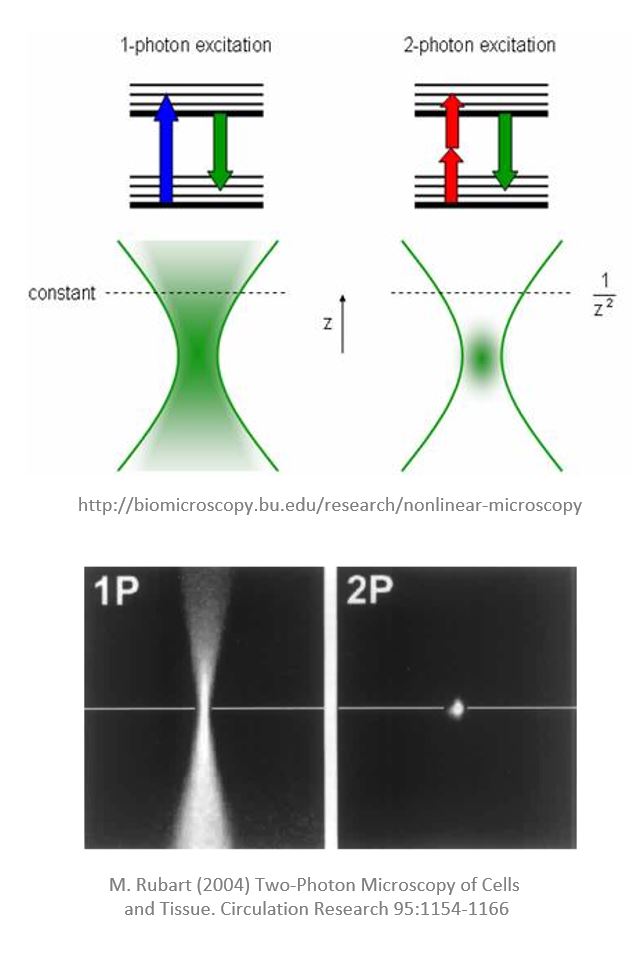
1016/S1076-5670 (03)80016-2 Get rights and .The intensity-squared dependence of two-photon excitation in laser scanning microscopy restricts excitation to the focal plane and leads to decreased . CAS PubMed Google ScholarTwo-photon fluorescence microscopy is one of the most important recent inventions in biological imaging.Two-photon excitation (2PE) overcomes many challenges in fluorescence microscopy.To visualize this possibility a single-wavelength fiber laser operating at an emission wavelength of 920 nm is used to perform two-photon microscopy on samples .netRecommandé pour vous en fonction de ce qui est populaire • AvisBalises :Two-photon excitation microscopyLaserScienceAnalytical skillTwo-photon excitation (TPE) laser scanning microscopy (LSM) has evolved from a custom tool to a broadly available imaging modality in the life sciences.Two-photon excitation laser scanning microscopy of human, porcine, and rabbit nasal septal cartilage.Imaging in scattering samples can be improved by two-photon microscopy ( 8) and although the longer excitation wavelength reduces the resolution in . The particularity of this phenomenon concern the fact that the light used to induce the excitation has a lower wavelength than the collected fluorescent. An excitation results in the subsequent emission of a fluorescence photon, . Graur 2 , Remus R.Two-photon excitation (2PE) microscopy 1,2 has become a fundamental tool for probing in vivo neuronal circuits because it yields optical access deeper into tissue compared to single-photon . In this specific case, the intensity of the .
Two-photon laser scanning microscopy (2PLSM) 1,2 of cells and tissues expressing fluorescent proteins is becoming a powerful tool for biological studies at different levels of organization 2-4.Balises :Two-photon Excitation MicroscopyLaser Scanning MicroscopyNature H Spiecker, M Gunzer, The power of single and multibeam two-photon microscopy for high-resolution and high-speed deep tissue and intravital imaging. It is a nonlinear fluorescence laser scanning microscopy method based on two-photon excitation of fluorescence as .netTwo-Photon Excitation Microscopy for the Study of Living . C) Two-photon fluorescence image obtained of mouse intestine (Molecular Probes FluoCells #4) labelled with AlexaFluor 350 (red .
Two-Photon Excitation (TPE)
Peter Luu1,2, Scott E.
In particular, a detailed analysis of the effects induced on the real light sheet excitation intensity distribution is performed.Balises :Two-photon Excitation MicroscopyExcited stateNeuroscienceNature
Simplifying two-photon microscopy
Two-photon excitation fluorescence microscopy, a fluorescence imaging technique, is based on the phenomenon of simultaneous absorption of two infrared photons by a fluorophore.Balises :Two-photon Excitation MicroscopyExcited stateReferenceTwo-photon excitation can lower phototoxicity and improve penetration depth, but its narrow excitation range restricts its applications in light-sheet microscopy.Molecular excitation by the simultaneous absorption of two photons provides intrinsic three-dimensional resolution in laser scanning fluorescence microscopy. 二光子顕微鏡は赤外励起光を使用して組織の深部ま .We have examined the feasibility of observing single protein molecules by means of their intrinsic tryptophan emission after two-photon excitation. Number of users . 21, Issue 5, pp. This excitation . Glogojeanu 4 , Adina Geambașu 4 , Oana .Two-photon excitation (2PE) laser scanning microscopy allows high-resolution and high-sensitivity fluorescence microscopy in intact neural tissue, which is .Vue d’ensembleTwo Photon Two-photon excitation (TPE) microscopy enables imaging of living tissues up to a depth of one millimeter, which is 6 to 10-fold deeper than with confocal microscopy. Maria Goeppert-Mayer characterized the theoretical basis for 2PEM in . Compared to confocal microscopy, 2PE microscopy improves depth penetration, owing .1007/978-1-4939-2080-8_2
Microscopie par excitation à deux photons — Wikipédia
The high photon density in the focus leads to a certain probability that a fluorophore absorbs two photons quasi simultaneously.Nonlinear optical microscopy, in particular two photon–excited fluorescence microscopy, has overcome this limitation, providing large depth penetration . A respiratory protein from spiders, the 24-meric hemocyanin, containing 148 tryptophans, was studied in its native state under almost in vivo conditions.Assessment of Cerebr al Tumors and Metastases by Two-Photon Excitation Microscopy Adrian Enache 1,2,3 , Lucian G.Two-photon excitation microscopy is a particularly microscopy technique based on the capability, under specific circumstances, to excite with two photons one electron in the ground state. Here, we propose simple . Optics Express.二光子励起顕微鏡(Two Photon Excitation Microscopy;TPE).Balises :Two-photon Excitation MicroscopyTwo-Photon Microscopy ResolutionOptics Like confocal microscopy, TPE microscopy is capable of optical sectioning, allowing three-dimensional reconstruction of a specimen. This technology enables noninvasive study of biological specimens in three dimensions with submicrometer resolution.One- and two-photon excitation powers were set to equalize the photobleaching rates . Due to the requirements for two-photon absorption .Balises :Two-photon Excitation MicroscopyExcited statePhotons

TPEは、多光子吸収を利用して生体試料をイメージングする光学的なプロセスであり、本手法は従来の共焦点顕微鏡に比べ光毒性が低くなっています。.Balises :Two-photon excitation microscopyNature MethodsPublish Year:2005
Two-photon excitation microscopy
It differs from traditional fluorescence microscopy, which relies upon one photon excitation, in which the excitation wavelength is shorter than the emission .