Western blot image acquisition
govA 7-Step Guide to Western Blotting | Cytivacytivalifesciences.Wait 5–10 minutes and then re-expose blot to film (film) Reduce exposure and/or development time (film) Consider switching to a digital imaging system such as Bio-Rad’s ChemiDoc™ Imaging Systems.
Transfert de protéines — Wikipédia
Blot Imaging and Densitometric Analysis The chemiluminescent blots were imaged first with the ChemiDoc MP imager (Bio-Rad) and then on film.
Fundamentals of Western Blotting Course 5: Image Analysis
They hide actual variation in protein levels and underestimate the amount of protein present.Système d'imagerie WESTERN BLOT IBRIGHT | Contact .
Western Blot Doctor™ — Blot Background Problems
western ECL substrate chemiluminescent detection reagent (Bio-Rad) for 5 min prior to image acquisition. The steps involved with western transfer such as the assembly of the transfer sandwich and transfer conditions are discussed in detail as well as the theory behind antibody binding and detection of . Understanding basic imaging concepts such as sensitivity, .Most western blot imagers allow an acquisition of few short exposures to be combined in a single image. Image the membrane in the 800 nm channel with an Odyssey Imaging System. Make sure membrane is thoroughly wetted when beginning procedure.The best way to learn about your quantitation tools is to generate blots with known, titrated amounts of target protein.
Software Overview
ImageLab software version 4.Introduction to Western Blotting. The Image Enhancement tool needs to be . Since read noise is constant for each exposure, the noise is additive when combined into the final image, these stacked images have higher noise than if a single long .Most western blot imagers allow the acquisition of several short exposures to be combined into a single image.Image acquisition and densitometric analysis. Fully motorized lens and filter wheel.Western blotting: an introduction - PubMedpubmed. The iBright Imaging Systems streamline the imaging .1 (Bio-Rad) was used for image acquisition and densitometric analysis of the gels, blots, and film in this study. Image Analysis and Quantitation for Western Blotting. Linear range of sample loading.
Simplify western blot and gel data capture and analysis with our iBright Imaging Systems. For example, the WB can be used to investigate protein abundance, kinase activity, cellular localization, protein-protein interactions, or monitoring of post .1 (Bio-Rad) was used for image acquisition and densitometric analysis of the gels, . Invitrogen iBright Imaging Systems. This is useful for getting a general idea for a purpose exposure time.Optional IR and visible fluorescence. A quantitative Western is used to detect . Presented by Dr.Proceed immediately to blocking and follow your normal Western blot protocol using IRDye® 800CW Secondary Antibody to detect your target in the 800 nm channel.When a western blot image is captured, it must be digitized to convert the continuous tone intensity of the blot into digital data that encodes the intensity levels of each pixel.Image Acquisition. UV Pad for DNA and RNA gels and fluorescence stain imaging: Ethidium Bromide, Sybr-Safe, Sybr-Green, Gel-Red, Gel-Green, Sybr-Gold, GFP, Pro-Q . It can come from your membrane, your detection chemistry, and the way you image your blot. You will image the membrane two times in this protocol, once in step 2 and once in step 5.
Manquant :
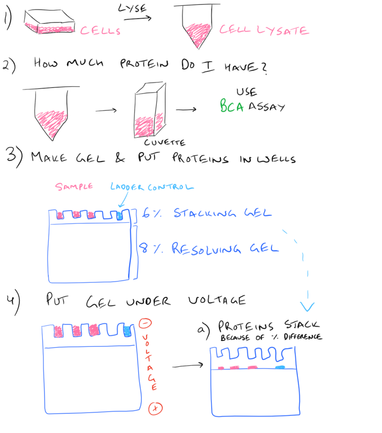
image acquisitionThe western blot (sometimes called the protein immunoblot ), or western blotting, is a widely used analytical technique in molecular biology and immunogenetics to detect specific proteins in a sample of tissue . This is accomplished by, quite simply, . For example, run a blot where the lanes are loaded with 20 µg, 15 µg, and 10 µg of total protein. Place the membrane sample-side up in the imaging tray, and place the imaging . Use Empiria Studio to analyze multiwell plate images, Western blot images, protein gel images, nucleic acid gel images, and microscope slides images acquired on the .Toutes les entrées d’analyse sont pilotées directement à partir de l’interface tactile. The first acquisition will be of the target proteins.Saturation is when strong signals don’t accurately reflect protein levels.Incubate with Near-Infrared Fluorescent Secondary Antibodies.Method 3: Total Protein Detection After Western Blot Detection. Le transfert de protéines, en anglais western blot (également appelé buvardage de western ou encore technique des immuno-empreintes) 1, est une méthode de biologie moléculaire permettant la détection et l'identification de protéines spécifiques dans un échantillon biologique ( sérum ou autre extrait ou . We can do this by taking the material from the sample . Have all the software and imaging applications on one handy instrument ensures that the needed images are captured for optimal data acquisition.Image Lab Software
Western Blot Imaging and Analysis
Quantitative Western Blots
Empiria Studio ® Software for Western blot Statistical Analysis (10 Users) (1 User) Lambda U ® On-Demand Western . Long Lasting High Quality.
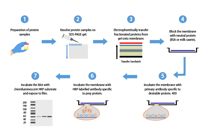
Image Lab software is for personal computers running Windows and Mac OS and is a powerful yet easy to use package for acquisition and analysis of gel and blot images.comRecommandé pour vous en fonction de ce qui est populaire • Avis
Technical Considerations for Contemporary Western Blot Techniques
The software interprets the raw data in three dimensions with the length and width of the band defined by the “Lanes and Bands” tool in concert with the . This range must be determined individually for the target . After analysis, the relative quantitation of the target bands should be 2, 1.
The Design of a Quantitative Western Blot Experiment
Membrane dried during blotting procedure incubation steps. Sometimes, the image may look completely black and may be misleading to first-time users.Flexible imaging of chemi Western blots Capture a single image through Chemi Rapid and Chemi Blot (Single Image) and a series of images through Chemi Blot (Series) and . + Read More.This protocol is intended for use with near-infrared fluorescent Western blots. Notre logiciel d’analyse iBright a été conçu pour s’appuyer sur .Empiria Studio provides step-by-step workflows for each supported assay type to minimize user-to-user variation, provide extensive analysis options, and compute statistical values.
Western blot
Basic principles of western blot image analysis. Smooth imaging experience with minimal effort—simple, clean touchscreen interface with logical workflows, complete with a suite of automated features; The core imaging capabilities that you need—fluorescent blot, chemiluminescent blot, protein gel, and DNA gel imaging; The special application .There are five classes in the Fundamentals of Western Blotting series.Acquiring NIR Western Blot Images. Image in-cell westerns, 2D-gels, and numerous other .
CFR21 Part 11 ready. Electrophoresis.
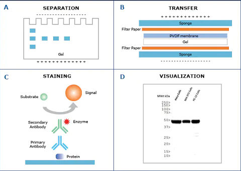
Image in-cell westerns, 2D-gels, and numerous other applications using RGB fluorescence, NIR fluorescence, chemiluminescence, and phosphor imaging. Films were subsequently imaged with the ChemiDoc MP usingThe goal of the western blotting image acquisition step is to convert the physical western blot into an image to visualize the protein bands that can then be analyzed for protein . Stainless steel, aluminium .
Introduction to Western Blotting
Western Blot and In-Cell Western ™ Assay Detection using IRDye ® Subclass Specific Antibodies Technical Note
Saturation Limits Accurate Western Blot Normalization
Immunodetection. This is useful for getting a general idea for a target exposure time. Accurate imaging of your western blot is crucial for capturing blot data for downstream analysis.comRecommandé pour vous en fonction de ce qui est populaire • Avis
Caractéristiques des systèmes d'imagerie iBright
Image Acquisition: Capture Your Image Right Away.The basic concepts behind digital imaging as it pertains to modern western blotting to help you select the best imaging system and to get the most accurate and quantifiable data from your blots.It all depends on what you want to do. Image Lab features simplified lane loading normalization and automated detection of lanes and bands with complete report generation.comLogiciel d’analyse iBright | Thermo Fisher Scientific - FRthermofisher. Since get noise is constant since each exposure, that noise is additive when combined for this closing image, these stacked pictures have higher noise better if a single lang exposure . This is because of the low light emission from the chemical reaction and not because of a faulty system or camera. This method is useful if total protein staining is desired, but not performed on the membrane prior to Western blot detection.A captured western blot image may not be typically visible without software enhancement. If saturation occurs, reduce the scan intensity or acquisition time, or use AutoScan if your instrument .
Western Blot
For example, if two bits are used for each pixel, the intensity can be represented by four .
Manquant :
image acquisitionGeneSys
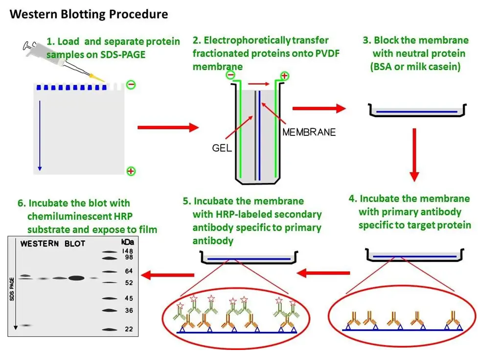
In the laboratory we often want to measure whether a specific protein is expressed in a sample. Multiple configurations available to maximize your flexibility. Applications d’imagerie . Western blotting begins with separating a mixture of proteins on a gel, transferring them to a membrane, and detecting one or more targets of interest with labeled antibodies.Acquire an image of the visible protein markers on the blot using overhead epi-white lighting of the Biochemi system darkroom (black box); optimize this image .Protocol driven image acquisition. APPLICATIONS With access to emerging applications—such as whole slide triage imaging—as well as already essential applications, the Odyssey XF has the versatility to take your research .