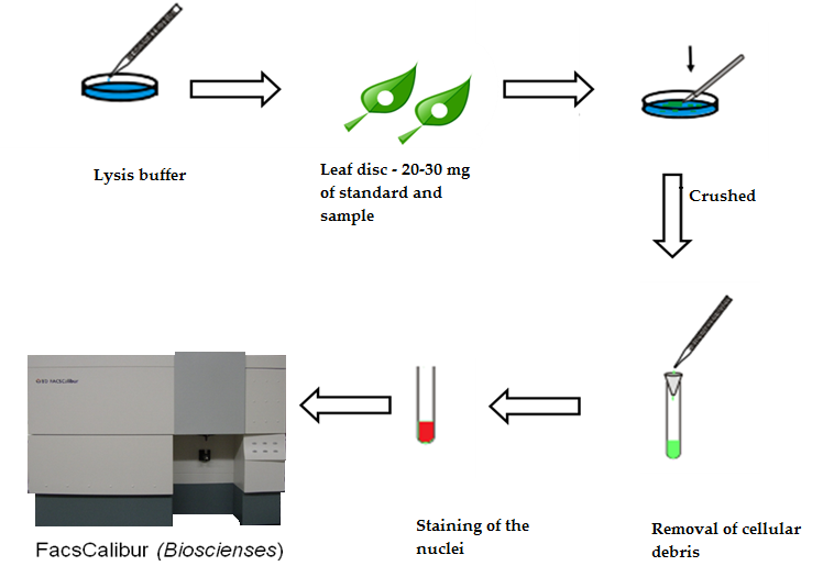
Flow cytometry preparation protocol
The following three points are important to consider: To prepare an ex vivo . It combines acoustic focusing technology coupled with traditional hydrodynamic focusing. If the aim is to detect only cell surface markers, .PBMC Preparation for Flow Cytometry **These protocols are meant to be modified with your experiment specifics in mind.Flow cytometry protocol. Continue as with suspension cultur e cells preparation protocol. Download Flow Cytometry Protocols Handbook. Single-cell suspensions are required for all flow cytometry . Positive staining with anti-CD16/32 on cells will show cells that . Not for use in diagnostic procedures. Flow Cytometry Protocol (Flow) IMPORTANT: Please refer to the APPLICATIONS section on the front page of product datasheet or product webpage to .
Flow cytometry (FACS) staining protocol (Cell surface staining)
Resources & Tools.
Cell Press: STAR Protocols
Flow Cytometry Guide
Sample preparation for flow cytometry involves several main steps, which can vary depending on the experiment.Sample preparation reagents for flow cytometry include cell surface staining, intracellular and transcription factor staining buffer sets, cell lysis assays, blocking reagents, and .
Preparation of Tissue Culture Cells for Flow Cytometry
Direct Immunofluorescence . For the preparation of single cells derived from . These protocols are designed for intracellular or cell . Flow cytometry measures these characteristics of . This section will yield . Optional: Block non-specific Fc-mediated interactions. Discover tips on finding the right fluorochrome for your experiment with our guides on .Sample preparation for flow cytometry involves several main steps, which can vary depending on the experiment.Flow cytometry. The solution is ready to add to the sample. EdU (5-ethynyl-2´-deoxyuridine) is a nucleoside analog to thymidine and is incorporated into DNA during active DNA synthesis. Pass cells from the tissue culture dish through the cell strainer to eliminate clumps and debris. The following flow cytometry staining protocols have been developed and optimized by R&D Systems Flow Cytometry Laboratory.This method was replaced by antibody-based detection of the nucleoside analog bromo-deoxyuridine (BrdU).
Flow Cytometry Sample Preparation Protocols
Download this handbook for information on protocols including: Sample preparation. Sample Preparation. Data Acquisition.Harvest cells from the serum/separation media interface using a pipet. Proceed to running samples on the flow cytometer.Key Flow Cytometry Protocol Steps. 還沒有帳戶嗎 ? 立即註冊. Last edited Mon 27 Mar 2023.
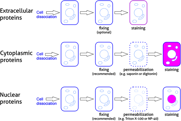
Flow Cytometry – BestProtocols® Page 2 of 4 Cell Preparation for Flow Cytometry Research Use Only For additional questions, please contact Technical Support at +1-888 . Stage 1 - Sample preparation . The Click-iT® EdU Flow Cytometry Assay Kits are novel alternatives to the BrdU assay. Discard supernatant and resuspend pellet in an . Reviewing the basics of flow cytometry, . The flow cytometer used in this protocol was the Invitrogen™ Attune™ NxT Flow Cytometer, equipped with 4 lasers (405 nm Violet, 488 nm Blue, 561 nm Yellow, and 637 nm Red) and 14 fluorescent detectors. Super Bright Staining Buffer protocol. Spectral Flow Cytometry Fundamentals . Solutions and Reagents. Sample preparation.mgdc3s6 February 7, 2018 4/10 4.Flow Cytometry is used for research applications such as immunophenotyping, DNA studies, cell cycle analysis, and fluorescence-activated cell sorting (FACS).Alternatively, mash tissue between the frosted ends of two microscope slides using 10 mL of Flow Cytometry Staining Buffer. Add ColorWheel ® finishing buffer to the ColorWheel ® antibody/dye mixture and incubate at 27 °C/room temperature for 30 minutes. Hence, should the reader wish to use alternative antibodies or cell types, experimental details specific to those parameters should be validated as described in . Single-cell suspensions are required for all . General procedure for detecting intracellular or extracellular proteins in flow cytometry. Substituting different products is not recommended.Preparation of cells stored in liquid nitrogen. Resuspend cells in cold PBS/BSA buffer and transfer them to a 15 ml conical centrifuge tube.Our success in developing a replicable protocol supported the development of a standardized workflow for rigorous selection and evaluation of antibodies and sample preparation conditions for flow cytometry experiments (Figure 6). The following three points are important to consider: To prepare an ex vivo cell population, freshly dissected .Accutase
Cell Preparation for Flow Cytometry
Any cell population that can be made into a single cell suspension can be assessed by flow cytometry.Prepare cells as described in BestProtocols: Cell Preparation Protocols for Flow Cytometry.Preparation of Cells for Flow Cytometry.Specifically, validation of a flow cytometry protocol requires an investigation into the relationship between sample preparation and obtained signal, which can only be defined by proper controls. Cell Preparation for Flow Cytometry Protocols (Invitrogen eBioscience reagents) Red . Centrifuge at 300-400 g for 5 min at 4 o C.Reliable Protocols for Flow Cytometry Analysis of Intracellular Proteins in Pluripotent Stem Cell Derivatives: A Fit-For-Purpose Approach - PMC. We will then transfer our single-cell suspension into 96-well plates, test tubes, or polystyrene . Hamburger Menu Button. Place harvested cells in a 15 ml conical centrifuge tube. Flow cytometry protocol.This protocol provides instructions on how to assemble Flex-T™ monomers into tetramers when needed.
PBMC Preparation for Flow Cytometry
There are four steps in most flow cytometry protocols: Sample Preparation.Because of its speed and ability to scrutinize at the single-cell level, flow cytometry offers the cell biologist the statistical power to rapidly analyze and characterize millions of cells. Place a cell strainer on top of a 15- or 15-mL conical tube. This is necessary when working with neutrophils, monocytes, macrophages, B-cells, natural killer cells, and some T cell subsets. Note: This protocol is intended for use with the specific products mentioned within it. Adjust the volume to 10 ml with PBS/BSA.Flow cytometry protocol | Abcam.There are four steps in most flow cytometry protocols: Sample Preparation; Blocking; Antibody Incubation; Data Acquisition; Related Products; Sample Preparation. Discard supernatant and resuspend pellet to a final concentration of at least 1 x 10 7 cells/ml with cold (4 o C) PBS . Preparation of Tissue Culture Cells Stored in Liquid Nitrogen 44. Resuspend cells in an appropriate volume of staining buffer, with care to avoid concentrations that will result in formation of cell aggregates. Single cells must be suspended at a density of 105 . If the aim is to detect only cell surface markers, the workflow involves processing the sample into a single-cell suspension, followed by blocking, immunostaining, and washing. Invitrogen eBioscience Resources—Selection guides, Best Protocols, product performance and more. Prepare PBS/BSA.
Live/dead staining with a .The ColorWheel ® Flow Cytometry Reagent Preparation protocol takes less than 5 minutes of hands-on time for a simplified reagent preparation for flow cytometry .There are four steps in most flow cytometry protocols: Sample Preparation; Blocking; Antibody Incubation; Data Acquisition; Sample Preparation.
Sample preparation for flow cytometry. Centrifuge cell suspension at 300-400 x g for 4-5 minutes at 2 . Phenotypic analysis. Proper Controls. Flow cytometry is a laboratory technique that is used to analyze cells. 產品 Antibodies Oligos, Primers, Probes and Genes TaqMan Real-Time PCR Assays Cell Culture Media Chemicals Western Blot Products Chromatography . Finally, it includes details on how to utilize a fluorophore-conjugated streptavidin to detect antigen-specific T cells .Use the following sample preparation protocols based on your appropriate starting materials: Preparation of suspension culture cells Preparation of adherent .Get flow cytometry protocols for cell preparation, red blood cell lysis, staining cells, compensation beads, viability and cell proliferation.Flow Cytometry Protocols. For Research Use Only.Immunofluorescent Staining of Intracellular Cytokines for Flow Cytometric Analysis.IMPORTANT: Please see the product-specific Flow Cytometry protocol on the product webpage for appropriate fixation and permeabilization conditions, and recommended antibody dilution.

BestProtocols: Cell Preparation for Flow Cytometry Protocols
Wash 1-3 times as described throughout this protocol.BestProtocols: Cell Preparation for Flow Cytometry Protocols.General protocols for flow cytometry.Protocols for Preparing Cells for Flow Cytometry 44.What is flow cytometry? Cytometry is the measurement of physical or chemical characteristics of cells or particles.io | https://dx.
The chapters in this book cover cytometry basics such as lasers for cytometry, metrics that can be used to evaluate spillover spreading, and the process of panel design and . First, we should harvest our cells or tissue and prepare a single-cell suspension. Resources & Tools.
Flow Cytometry Protocol (Flow)
Thaw cells rapidly in a 37 o C water bath. This workflow outlines major steps required to establish the fit-for-purpose of an antibody and protocol for . Any cell population that . Staining cells with a No-Lyse protocol. Centrifuge at 300-400 g for 5 minutes at room temperature. Step 3 in the ColorWheel ® Flow Cytometry Reagent Preparation protocol.
BestProtocols: Staining Cell Surface Targets for Flow Cytometry
It also covers how to exchange the dummy peptide in the MHC groove for the researcher's peptide of interest with an ultraviolet (UV) lamp.Fluorophore and reagent selection guide for flow cytometry. Carefully remove cells from liquid nitrogen storage.
Flow cytometry protocol
Direct Immunofluorescence Staining of Mononuclear Cells.Technical resources / / Flow cytometry protocol.